. 2010 Aug 17;5(8):e12220.
doi: 10.1371/journal.pone.0012220.

Role of mast cells in inflammatory bowel disease and inflammation-associated colorectal neoplasia in IL-10-deficient mice

Affiliations

Role of mast cells in inflammatory bowel disease and inflammation-associated colorectal neoplasia in IL-10-deficient mice

Maciej Chichlowski et al. PLoS One. .

Abstract

Background: Inflammatory bowel disease (IBD) is hypothesized to result from stimulation of immune responses against resident intestinal bacteria within a genetically susceptible host. Mast cells may play a critical role in IBD pathogenesis, since they are typically located just beneath the intestinal mucosal barrier and can be activated by bacterial antigens.

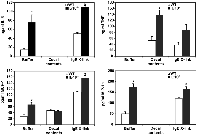
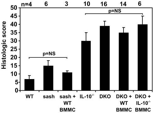
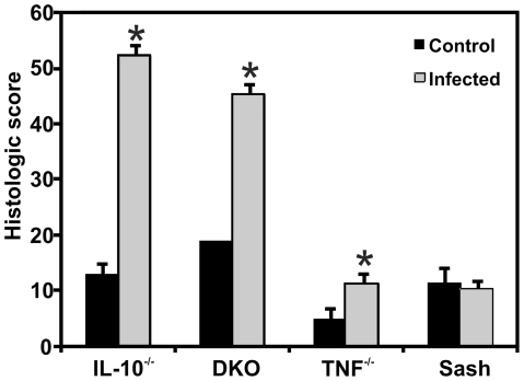
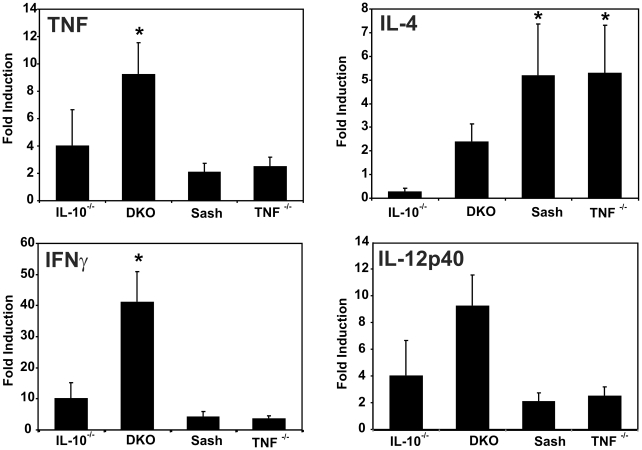
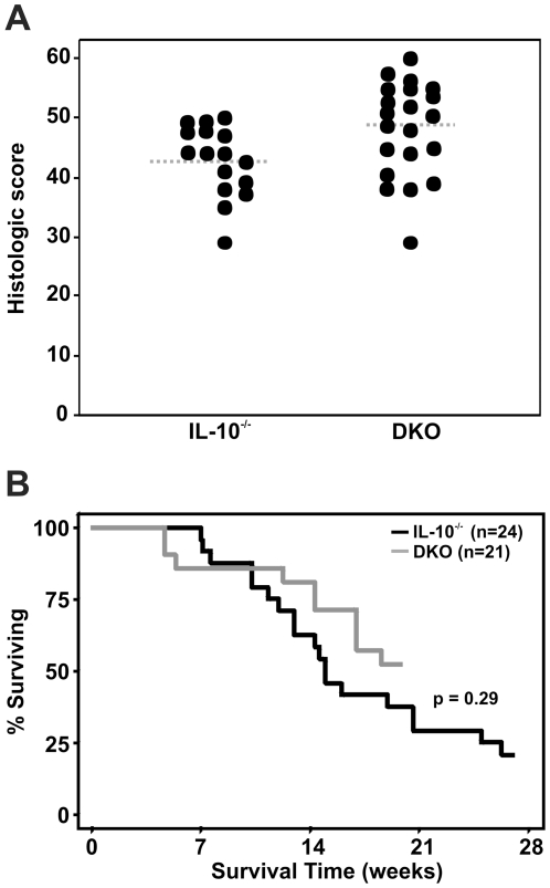
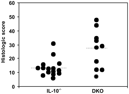
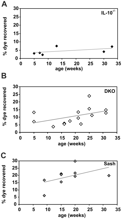
Methodology/principal findings: This study investigated effects of mast cells on inflammation and associated neoplasia in IBD-susceptible interleukin (IL)-10-deficient mice with and without mast cells. IL-10-deficient mast cells produced more pro-inflammatory cytokines in vitro both constitutively and when triggered, compared with wild type mast cells. However despite this enhanced in vitro response, mast cell-sufficient Il10(-/-) mice actually had decreased cecal expression of tumor necrosis factor (TNF) and interferon (IFN)-gamma mRNA, suggesting that mast cells regulate inflammation in vivo. Mast cell deficiency predisposed Il10(-/-) mice to the development of spontaneous colitis and resulted in increased intestinal permeability in vivo that preceded the development of colon inflammation. However, mast cell deficiency did not affect the severity of IBD triggered by non-steroidal anti-inflammatory agents (NSAID) exposure or helicobacter infection that also affect intestinal permeability.

Conclusions/significance: Mast cells thus appear to have a primarily protective role within the colonic microenvironment by enhancing the efficacy of the mucosal barrier. In addition, although mast cells were previously implicated in progression of sporadic colon cancers, mast cells did not affect the incidence or severity of colonic neoplasia in this inflammation-associated model.


Conflict of interest statement

Competing Interests: The authors have declared that no competing interests exist.

Figures

Figure 1
Figure 1. BMMC cytokine production after stimulation with IgE or enteric bacteria.
BMMC were stimulated for 4 hrs, then media was harvested for cytokine analysis via Luminex bead-based fluorescent immunoassays. Il10 / BMMC make more IL-6, MCP-1, and MIP-1α constitutively (buffer treatment) and following IgE stimulation than WT BMMC (p<0.05). Exposure to enteric bacteria in cecal contents triggers a different pattern of cytokine expression, with higher TNF production by Il10 −/− vs. WT BMMC. Data shown is the mean ± SEM for 3–5 independent experiments. * indicates p≤0.05 vs. WT.
Figure 2
Figure 2. Mast cells do not affect the severity of colitis triggered by piroxicam.
Mice of the indicated genotypes ± reconstitution with WT or Il10 −/− BMMC received 200 ppm piroxicam ×7 days to trigger the onset of colitis. Tissue was harvested for histologic analysis 16 d later. WT or sash mice ± reconstitution with WT BMMC did not develop colitis in this study. Mice deficient in IL-10 uniformly developed colitis, however the presence or absence of mast cells and whether or not the mast cells could produce IL-10 did not affect the severity of inflammation.
Figure 3
Figure 3. Mast cells do not affect early colitis severity in Helicobacter-infected Il10 −/− mice.
Both IL-10-deficient and DKO mice exhibited severe colonic inflammation when infected with H. rodentium and H. typhlonius for 30 d (n = 3 for Ill0 −/− and n = 4 for DKO mice) compared to Tnf −/− and sash mice (n = 4 each) (p<0.001 for both Il10 −/− and DKO vs. Tnf −/− and sash). * indicates p<0.01, comparing control (non-infected) and infected mice of a given genotype.
Figure 4
Figure 4. Mast cells modulate pro-inflammatory cytokine mRNA expression in inflamed cecal tissues.
Cytokine mRNA expression in cecal tissue of mice of the indicated genotypes harvested after 5 weeks of infection with H. rodentium and H. typhlonius was measured by real-time PCR, normalized to β-actin and expressed as fold-induction relative to non-infected cecal tissue of the same genotype. Bars represent 5–6 mice per genotype. * indicates p<0.05 compared to Il10 −/− mice. Infected DKO mice have increased expression of Th1 cytokines (TNF, IFN-γ) and a trend towards increased Th2 (IL-4) and Th17-inducing cytokines (IL-12/23p40) compared with mast cell-sufficient Il10 −/− mice.
Figure 5
Figure 5. Mast cells do not affect long-term colitis severity in Helicobacter-infected Il10 −/− mice.
Il10 −/− mice with (n = 21) and without (n = 24) genetic deficiency of mast cells were infected with H. rodentium and H. typhlonius and monitored for up to 27 wks. DKO mice had a small increase in histologic score compared with mice deficient in IL-10 alone (A; p = 0.03), but there was no difference in survival between DKO and mast cell-sufficient Il10 −/− mice (B).
Figure 6
Figure 6. Colon histology and neoplasia in Helicobacter-infected and non-infected Il10 −/− and DKO adult mice.
Helicobacter-infected Il10 −/− (A, B) and DKO mice (C, D) uniformly exhibited mucosal hyperplasia with prominent inflammatory infiltrates. Examples of neoplastic lesions seen in the long-term study are shown. The arrow in D indicates malignant glands that have invaded into the serosa. Bar represents 1 mm in the large panels and 100 µm in the insets.
Figure 7
Figure 7. Severity of spontaneous IBD in specific pathogen-free Il10 −/− and DKO mice.
Il10 −/− mice on the C57BL/6 background rarely developed spontaneous colitis by 36 weeks of age when housed under clean conditions, free from specific pathogens including Helicobacter spp. (mean histologic scores ± SEM = 13±2; n = 15). In contrast, DKO mice that lacked mast cells frequently developed spontaneous colitis (mean histologic scores = 27±4; n = 10; p = 0.02), which was evident at an earlier age (31±2 wks vs. 36±1 wks; p = 0.02) compared with Il10 −/− mice.
Figure 8
Figure 8. Permeability of the intestinal barrier in IL-10−/− and DKO mice.
Mice of the indicated ages and genotypes were gavaged with phenol red and the amount of dye absorbed was determined by spectrophotometric measurement of urine. Each point shown represents the average of 2–4 mice tested in a single experiment. Mast cell-deficient DKO and sash mice had significantly increased dye recovery compared with Il10 −/− mice (p = 0.02 for differences in data point elevation via linear regression analysis). The total numbers of mice tested were: Il10 −/− (n = 14), DKO (n = 18), and sash (n = 22).

References

    1. Chichlowski M, Hale LP. Bacterial-mucosal interactions in inflammatory bowel disease—an alliance gone bad. Am J Physiol Gastrointest Liver Physiol. 2008;295:G1139–1149. - PMC - PubMed
    1. Sollid LM, Johansen F-E. Animal Models of Inflammatory Bowel Disease at the Dawn of the New Genetics Era. PLoS Med. 2008;5:e198. - PMC - PubMed
    1. Inaba Y, Ashida T, Ito T, Ishikawa C, Tanabe H, et al. Expression of the antimicrobial peptide alpha-defensin/cryptdins in intestinal crypts decreases at the initial phase of intestinal inflammation in a model of inflammatory bowel disease, IL-10-deficient mice. Inflammatory Bowel Diseases. 2010;9999:NA. - PubMed
    1. Kühn R, Löhler J, Rennick D, Rajewsky K, Müller W. Interleukin-10-deficient mice develop chronic enterocolitis. Cell. 1993;75(2):263–274. - PubMed
    1. Hale LP, Gottfried MR, Swidsinski A. Piroxicam treatment of IL-10-deficient mice enhances colonic epithelial apoptosis and mucosal exposure to intestinal bacteria. Inflamm Bowel Dis. 2005;11:1060–1069. - PubMed

Publication types

MeSH terms