. 2010 Aug 17;5(8):e12228.
doi: 10.1371/journal.pone.0012228.

bFGF regulates PI3-kinase-Rac1-JNK pathway and promotes fibroblast migration in wound healing

Affiliations

bFGF regulates PI3-kinase-Rac1-JNK pathway and promotes fibroblast migration in wound healing

Shigeyuki Kanazawa et al. PLoS One. .

Abstract

Fibroblast proliferation and migration play important roles in wound healing. bFGF is known to promote both fibroblast proliferation and migration during the process of wound healing. However, the signal transduction of bFGF-induced fibroblast migration is still unclear, because bFGF can affect both proliferation and migration. Herein, we investigated the effect of bFGF on fibroblast migration regardless of its effect on fibroblast proliferation. We noticed involvement of the small GTPases of the Rho family, PI3-kinase, and JNK. bFGF activated RhoA, Rac1, PI3-kinase, and JNK in cultured fibroblasts. Inhibition of RhoA did not block bFGF-induced fibroblast migration, whereas inhibition of Rac1, PI3-kinase, or JNK blocked the fibroblast migration significantly. PI3-kinase-inhibited cells down-regulated the activities of Rac1 and JNK, and Rac1-inhibited cells down-regulated JNK activity, suggesting that PI3-kinase is upstream of Rac1 and that JNK is downstream of Rac1. Thus, we concluded that PI3-kinase, Rac1, and JNK were essential for bFGF-induced fibroblast migration, which is a novel pathway of bFGF-induced cell migration.


Conflict of interest statement

Competing Interests: The authors have declared that no competing interests exist.

Figures

Figure 1
Figure 1. Number of the cells treated with each concentration of mitomycin-C.
(A) We counted the number of fibroblasts in a microscopic counting chamber before or after treated with each concentration of mitomycin-C (0, 1, 5 or 10 µg/ml) for 24 h. Mitomycin-C at 0 or 1 µg/ml did not block cell proliferation. Mitomycin-C at 5 µg/ml completely blocked cell proliferation, while Mitomycin-C at 10 µg/ml was indicative of cellular cytotoxicity. (B) We counted the number of fibroblasts before or after treated with 0 or 5 µg/ml mitomycin-C in the presence of 100 ng/ml bFGF. Mitomycin-C at 5 µg/ml blocked bFGF-induced fibroblast proliferation. Data are mean ± s.e.m. of five independent experiments. **P<0.01, as compared with the control group (t test).
Figure 2
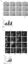
Figure 2. Effect of bFGF on fibroblast migration.
(A) Wound healing assay in the fibroblasts treated with the indicated concentrations of bFGF in the presence of 5 µg/ml mitomycin-C. This photograph shows the wounded cell monolayers at 0, 12, and 24 h after wounding in the absence or presence of bFGF. The line indicates the wound edge at the start of the experiment (0 h). Bar  = 500 µm. (B) Analysis of the migration rate is expressed as migration distance/time (µm/h). (C) Immunocytochemistry of the cells at the margin of the scratch wound with rhodamine-conjugated phalloidin in the presence of 5 µg/ml mitomycin-C. This photograph shows the F-actin of the cell at 0, 1, 6, and 12 h after wounding with or without 100 ng/ml bFGF. The bFGF-treated fibroblasts developed lamellipodial extension (arrowhead). Bar  = 50 or 10 µm (D) Lamellipodial extension was quantified by summing the length of the outer margins of all lamellipodium in individual cells, and was expressed as a proportion of the total perimeter for each cell (lamellipodial index). Data are mean ± s.e.m. of five independent experiments. **P<0.01, as compared with the control group (t test).
Figure 3
Figure 3. Activities of RhoA, Rac1, and Cdc42 by bFGF-stimulation.
(A)(B)(C) Activities in pull-down assays for RhoA, Rac1, and Cdc42 were analyzed at 15, 30, and 60 min after 100 ng/ml bFGF-stimulation in the presence of 5 µg/ml mitomycin-C. Densitometry for RhoA, Rac1, and Cdc42-GTP was normalized to the amount of total RhoA, Rac1, and Cdc42. Bradykinin treatment (100 ng/ml) for 10 min was performed as a positive control of Cdc42 activation. The results are presented as fold change as compared with fibroblasts in the absence of bFGF. Data are mean ± s.e.m. of three independent experiments. **P<0.01, as compared with the control group (t test). brad. =  bradykinin.
Figure 4
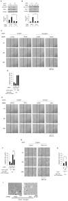
Figure 4. Rac1, but not RhoA, was essential for bFGF-induced fibroblast migration.
(A)(B) Primary rat fibroblasts were transfected with 10 nM RhoA, 10 nM Rac1 siRNA or 10 nM nonspecific control pool siRNA. Pull-down assays for RhoA or Rac1 in the presence of 5 µg/ml mitomycin-C were performed after 48 h of siRNA treatment, using an immunoblot of lysates for GAPDH to verify equal protein loading. (C)(E) Wound healing assay containing 5 µg/ml mitomycin-C in fibroblasts transfected with 10 nM RhoA, 10 nM Rac1 siRNA or 10 nM nonspecific control pool siRNA in the presence or absence of bFGF. Bar = 500 µm. (D)(F)(H) Analysis of the migration rate of the RhoA, Rac1-inhibited cells, or the active Rac1-taransfected cells was performed. (G) Wound healing assay treated with 5 µg/ml mitomycin-C in fibroblasts transfected with 0.15 µg/cm2 of plasmid DNA (Rac1-61L) or empty vector as a control in the absence of bFGF. (I) These images show the cell shape of the RhoA or Rac1-inhibited cells at 24 h after wounding in the presence of 100 ng/ml bFGF. Bar = 50 µm. Data are mean ± s.e.m. of five independent experiments. *P<0.05, **P<0.01, as compared with the control group (t test).
Figure 5
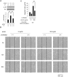
Figure 5. Effect of inhibition of PI3-kinase on fibroblast migration.
(A) The activity of Akt with or without 10 µM LY294002 was analyzed by immunoblotting at 30 min after 100 ng/ml bFGF-stimulation in the presence of 5 µg/ml mitomycin-C. Densitometry for p-Akt was normalized to the amount of Akt. The results are presented as fold change as compared with the fibroblasts in the absence of bFGF. (B) Wound healing assay containing 5 µg/ml mitomycin-C in fibroblasts treated with or without 10 µM LY294002 in the presence or absence of 100 ng/ml bFGF. Bar  = 500 µm. (C) Analysis of the migration rate of PI3-kinase-inhibited cells was performed. Data are mean ± s.e.m. of five independent experiments. **P<0.01, as compared with the control group (t test).
Figure 6
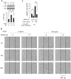
Figure 6. Effect of inhibition of JNK on fibroblast migration.
(A) The activity of JNK with or without 10 µM SP600125 was analyzed by immunoblotting at 30 min after 100 ng/ml bFGF-stimulation in the presence of 5 µg/ml mitomycin-C. Densitometry for p-JNK was normalized to the amount of JNK. The results are presented as fold change as compared with fibroblasts in the absence of bFGF. (B) Wound healing assay containing 5 µg/ml mitomycin-C in fibroblasts treated with or without 10 µM SP600125 in the presence or absence of 100 ng/ml bFGF. Bar  = 500 µm. (C) Analysis of the migration rate of JNK-inhibited cells was performed. Data are mean ± s.e.m. of five independent experiments. *P<0.05, **P<0.01, as compared with the control group (t test).
Figure 7
Figure 7. PI3-kinase contributed to bFGF-induced Rac1 and JNK activation.
(A)(D) The activities of Rac1 or JNK in lysates of fibroblasts treated with or without 10 µM LY294002 were analyzed at 15 min after 100 ng/ml bFGF-stimulation in the presence of 5 µg/ml mitomycin-C. (B) The activity of JNK in lysates of fibroblasts transfected with 10 nM Rac1 siRNA or 10 nM nonspecific control pool siRNA was analyzed at 15 min after 100 ng/ml bFGF-stimulation in the presence of 5 µg/ml mitomycin-C. (C) The activity of JNK without bFGF in lysates of fibroblasts transfected with 0.15 µg/cm2 of plasmid DNA (Rac1-61L) or empty vector as a control was analyzed in the presence of 5 µg/ml mitomycin-C. Data are mean ± s.e.m. of three independent experiments. *P<0.05, **P<0.01, as compared with the control group (t test).
Figure 8
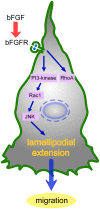
Figure 8. Signal transduction pathway of bFGF-stimulated fibroblast migration.
Activated FGFR stimulates PI3-kinase. Activation of PI3-kinase leads to up-regulation of Rac1, followed by the activation of JNK, resulting in formation of lamellipodial extension.

Similar articles

Cited by

References

    1. Wagner W, Wehrmann M. Differential cytokine activity and morphology during wound healing in the neonatal and adult rat skin. J Cell Mol Med. 2007;11:1342–1351. - PMC - PubMed
    1. Lauffenburger DA, Horwitz AF. Cell migration: a physically integrated molecular process. Cell. 1996;84:359–369. - PubMed
    1. Webb DJ, Parsons JT, Horwitz AF. Adhesion assembly, disassembly and turnover in migrating cells – over and over and over again. Nat Cell Biol. 2002;4:E97–E100. - PubMed
    1. Ridley AJ, Schwartz MA, Burridge K, Firtel RA, Ginsberg MH, et al. Cell migration: integrating signals from front to back. Science. 2003;302:1704–1709. - PubMed
    1. Smith LG, Li R. Actin polymerization: riding the wave. Curr Biol. 2004;14:R109–111. - PubMed

Publication types

MeSH terms

Substances