. 2010 Nov;31(11):1251-60.
doi: 10.1002/humu.21350.

Molecular mechanisms leading to null-protein product from retinoschisin (RS1) signal-sequence mutants in X-linked retinoschisis (XLRS) disease

Affiliations

Molecular mechanisms leading to null-protein product from retinoschisin (RS1) signal-sequence mutants in X-linked retinoschisis (XLRS) disease

Camasamudram Vijayasarathy et al. Hum Mutat. 2010 Nov.

Abstract

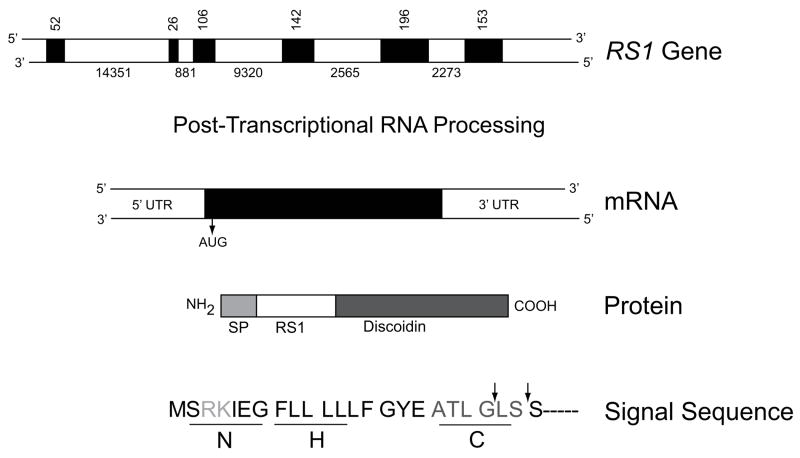
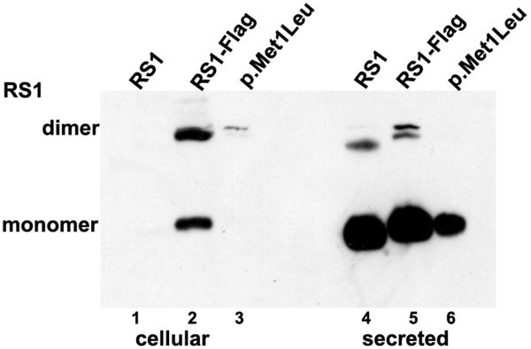
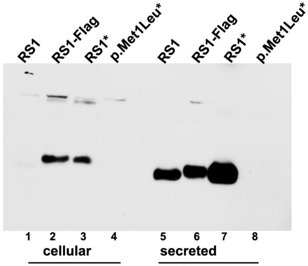
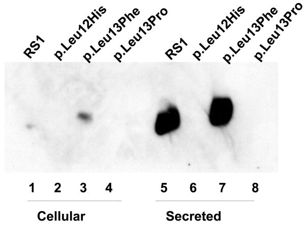
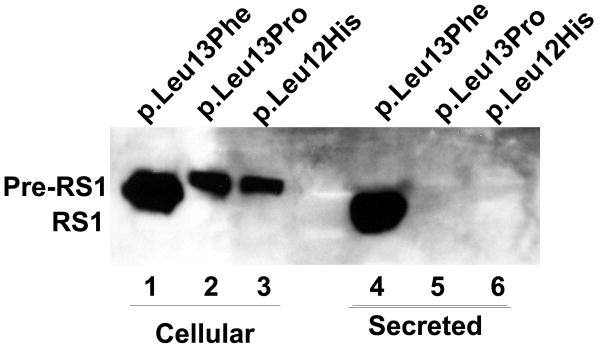
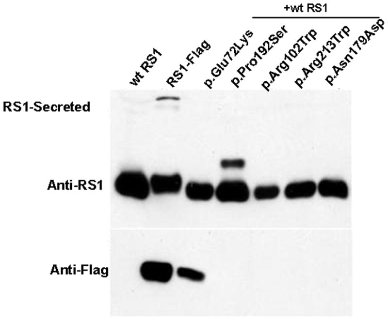
Retinoschisin (RS1) is a cell-surface adhesion molecule expressed by photoreceptor and bipolar cells of the retina. The 24-kDa protein encodes two conserved sequence motifs: the initial signal sequence targets the protein for secretion while the larger discoidin domain is implicated in cell adhesion. RS1 helps to maintain the structural organization of the retinal cell layers and promotes visual signal transduction. RS1 gene mutations cause X-linked retinoschisis disease (XLRS) in males, characterized by early-onset central vision loss. We analyzed the biochemical consequences of several RS1 signal-sequence mutants (c.1A>T, c.35T>A, c.38T>C, and c.52G>A) found in our subjects. Expression analysis in COS-7 cells demonstrates that these mutations affect RS1 biosynthesis and result in an RS1 null phenotype by several different mechanisms. By comparison, discoidin-domain mutations generally lead to nonfunctional conformational variants that remain trapped inside the cell. XLRS disease has a broad heterogeneity in general, but subjects with the RS1 null-protein signal-sequence mutations are on the more severe end of the clinical phenotype. Results from the signal-sequence mutants are discussed in the context of the discoidin-domain mutations, clinical phenotypes, genotype-phenotype correlations, and implications for RS1 gene replacement therapy.


Figures

Figure 1
Figure 1
Schematic structure of the RS1 gene, mRNA, and protein products. RS1 is encoded on the minus strand of the X chromosome at Xp22.2–p22.1 covering 32.43 kb from 18600150 to 18567724. Exons are indicated by filled boxes, with numbers indicating the size of the exons and introns in nucleotides. The primary RNA transcript encoding both exons and introns undergoes post-transcriptional RNA splicing to remove introns and generate mRNA (NM_000330.3*) which is translated into a 224 amino-acid protein (NP_000321.1). The functional domains of RS1 are 1) a signal peptide (SP), 2) RS1, and 3) the discoidin domains. The signal sequence guides the translocation of nascent RS1 from the endoplasmic reticulum (the site of synthesis) to external leaflet of the plasma membrane, during which signal sequence is cleaved by signal peptidase to generate mature protein with characteristic RS1 and a highly conserved discoidin domain. The different subdomains of RS1 signal sequence are 1) the positively charged N region at the amino terminal end which mediates translocation, 2) the hydrophobic core (H) required for targeting and membrane insertion and 3) a polar “C” region that determines the site of recognition and cleavage by signal peptidase. The arrows indicate the sites at which the signal peptide is cleaved. *The numbering follows GenBank NCBI Reference Sequence: NM_000330.3. Nucleotide 1 is A of the ATG initiation codon (CDS 36–710).
Figure 2
Figure 2
Fundus photographs and non-invasive optical coherence tomograms (OCT) through a horizontal section of both eyes of 13-y/o III.4 and 45-y/o II.2, both with the c.52G>A mutation of the RS1 gene. III.4 has classical foveal schisis prototypical of XLRS, while II.2 shows abnormal RPE pigmentation and atrophic retinal changes in the macular area of the right eye and the periphery of the left eye. Atrophic macular thinning is quite evident by the OCT scans. II.2 also presents a large bullous peripheral schisis, as seen in patients with a severe XLRS phenotype.
Figure 3
Figure 3
Electroretinogram depicting the rod- and cone-driven responses of the right eyes of two related males affected by XLRS (c.52G>A) and from a representative normal control subject. The rod-driven b-wave is relatively preserved in the 13-y/o III.4, but the 45-y/o-uncle II.2 shows remarkable reduction of b-wave amplitude, with the typical XLRS electronegative ERG waveform. The cone-mediated responses are overall normal in the young III.4 male, but are delayed and reduced in amplitude in the middle-aged II.2.
Figure 4
Figure 4
Effect of AUG alteration to UUG (p.Met1Leu) on RS1 mRNA and protein. (A) WT RS1 and p.Met1Leu mutants were transiently expressed in COS-7 cells. At 72 h post-transfection cultures were harvested and cellular and secreted fractions were analyzed for RS1 protein by immunoblotting using anti-RS1 rabbit polyclonal antibody raised against the N terminus amino-acid residues 24–37 of RS1. Both WT (AUG initiation codon) and Mut (UUG initiation codon) cells expressed RS1, which as expected, was mostly detected in the secreted fraction (lanes 4–6) as compared to the cellular bound RS1 (lanes 1–3). The results indicate that protein synthesis can be initiated at the UUG initiation codon. (B) RS1 expression in cells expressing plasmids encoding 5′ UTR immediately upstream of AUG (RS1*) or UUG (p.Met1Leu*) initiation codons. RS1 is profusely expressed in cells expressing the WT control plasmids RS1 (without 5′ UTR; lanes 1, 5) or RS1* (with 5′ UTR; lanes 3, 7). However, 5′ UTR immediately upstream of UUG initiation codon selectively abolished RS1 translation in cells expressing p.Met1Leu* (lanes 4, 8). (C) Detection of RS1 gene transcripts: total RNA isolated from cells expressing either WT or Mut expression plasmids without or with 5′ UTR was subjected to RT-PCR with RS1 cDNA specific primers and the products analyzed by agarose gel electrophoresis (semi-quantitative). Lane M is molecular size marker. The mRNA levels: lane 1, mouse retina; lane 3, the WT RS1* with 5′ UTR; lane 5, the mutant with 5′ UTR (p. Met1 Leu*); lane 7, p.Met1Leu without 5′ UTR. Parallel reactions were carried out in the absence of RT (even numbered lanes).
Figure 4
Figure 4
Effect of AUG alteration to UUG (p.Met1Leu) on RS1 mRNA and protein. (A) WT RS1 and p.Met1Leu mutants were transiently expressed in COS-7 cells. At 72 h post-transfection cultures were harvested and cellular and secreted fractions were analyzed for RS1 protein by immunoblotting using anti-RS1 rabbit polyclonal antibody raised against the N terminus amino-acid residues 24–37 of RS1. Both WT (AUG initiation codon) and Mut (UUG initiation codon) cells expressed RS1, which as expected, was mostly detected in the secreted fraction (lanes 4–6) as compared to the cellular bound RS1 (lanes 1–3). The results indicate that protein synthesis can be initiated at the UUG initiation codon. (B) RS1 expression in cells expressing plasmids encoding 5′ UTR immediately upstream of AUG (RS1*) or UUG (p.Met1Leu*) initiation codons. RS1 is profusely expressed in cells expressing the WT control plasmids RS1 (without 5′ UTR; lanes 1, 5) or RS1* (with 5′ UTR; lanes 3, 7). However, 5′ UTR immediately upstream of UUG initiation codon selectively abolished RS1 translation in cells expressing p.Met1Leu* (lanes 4, 8). (C) Detection of RS1 gene transcripts: total RNA isolated from cells expressing either WT or Mut expression plasmids without or with 5′ UTR was subjected to RT-PCR with RS1 cDNA specific primers and the products analyzed by agarose gel electrophoresis (semi-quantitative). Lane M is molecular size marker. The mRNA levels: lane 1, mouse retina; lane 3, the WT RS1* with 5′ UTR; lane 5, the mutant with 5′ UTR (p. Met1 Leu*); lane 7, p.Met1Leu without 5′ UTR. Parallel reactions were carried out in the absence of RT (even numbered lanes).
Figure 5
Figure 5
Immunoblot analysis of RS1 expression in cellular and secreted fractions of COS-7 cells expressing p.Leu12His and p.Leu13Pro mutants. (A) The WT RS1 and the p.Leu13Phe mutant with intact signal sequence hydrophobic core both profusely expressed RS1, which was processed and secreted into the culture medium (lanes 5, 7) with little retained in the cellular fractions (lanes 1, 3). The p.Leu12His (lanes 2, 6) and p.Leu13Pro mutants (lanes 6, 8), with a disrupted hydrophobic core, showed virtual lack of RS1 expression. (B) Proteasomal inhibition stabilizes RS1 protein in the mutants. COS-7 cells were transfected with RS1 expression plasmids one day before MG132 (10 μM for 6h); or its vehicle dimethyl sulfoxide (DMSO) was added, and the cells were grown in serum-supplemented medium. The precursor form of RS1 (224 aa) stabilized in the presence of MG132 is seen in the cellular fractions of the signal sequence mutants (lanes 1–3). In p.Leu13Phe the precursor is processed into mature form of RS1 (201 aa) and secreted into the medium (lane 4) but not in the p.Leu12His and p.Leu13Pro mutants due to a processing defect.
Figure 5
Figure 5
Immunoblot analysis of RS1 expression in cellular and secreted fractions of COS-7 cells expressing p.Leu12His and p.Leu13Pro mutants. (A) The WT RS1 and the p.Leu13Phe mutant with intact signal sequence hydrophobic core both profusely expressed RS1, which was processed and secreted into the culture medium (lanes 5, 7) with little retained in the cellular fractions (lanes 1, 3). The p.Leu12His (lanes 2, 6) and p.Leu13Pro mutants (lanes 6, 8), with a disrupted hydrophobic core, showed virtual lack of RS1 expression. (B) Proteasomal inhibition stabilizes RS1 protein in the mutants. COS-7 cells were transfected with RS1 expression plasmids one day before MG132 (10 μM for 6h); or its vehicle dimethyl sulfoxide (DMSO) was added, and the cells were grown in serum-supplemented medium. The precursor form of RS1 (224 aa) stabilized in the presence of MG132 is seen in the cellular fractions of the signal sequence mutants (lanes 1–3). In p.Leu13Phe the precursor is processed into mature form of RS1 (201 aa) and secreted into the medium (lane 4) but not in the p.Leu12His and p.Leu13Pro mutants due to a processing defect.
Figure 6
Figure 6
Sequence of eight highly conserved nucleotides at the boundary between an exon and an intron: the 5′ donor splice site (5′ ss) of eukaryotic mRNAs (Shapiro and Senapathy, 1987). Also shown are the sequences at the boundary between exon 1 and intron 1 in RS1 WT and c52 G>A mutant. Splicing is catalyzed by spliceosome, an RNA/protein complex consisting of small nuclear ribonucleoprotein particles (snRNPs) and SR family of splicing proteins (splicing factors with one or more RNA-recognition motif) through RNA–RNA and protein–protein interactions. snRNPs are composed of snRNAs (U1, U2, U4N6, and U5) each associated with 8 sm proteins. These snRNPs assemble on the conserved sequence motifs at three sites: U1 at exon–intron junction (5′ splice site), U2AF at intron–exon junction (3′ splice site) and U2 at branch point located 18–40 nucleotides upstream of the 3′ splice site.
Figure 7
Figure 7
RS1 RNA and protein analysis in cells expressing the WT or c.52G>A Mut minigene. (A) Cartoon of the mutant c.52G>A minigene used. The minigene without splice-site mutation served as control WT. (B) Immunoblot analysis of RS1 protein with anti-RS1 antibody detected RS1 protein both in the cellular and secreted fractions of the cells expressing the WT minigene. RS1 protein was not detected in the fractions derived from cells expressing the c.52G>A mutant minigene.
Figure 7
Figure 7
RS1 RNA and protein analysis in cells expressing the WT or c.52G>A Mut minigene. (A) Cartoon of the mutant c.52G>A minigene used. The minigene without splice-site mutation served as control WT. (B) Immunoblot analysis of RS1 protein with anti-RS1 antibody detected RS1 protein both in the cellular and secreted fractions of the cells expressing the WT minigene. RS1 protein was not detected in the fractions derived from cells expressing the c.52G>A mutant minigene.
Figure 8
Figure 8
Effect of missense mutations in the discoidin domain on RS1-protein expression and localization. (A) The missense mutants were transiently expressed in COS-7 cells and analyzed for RS1 expression. The WT RS1 was predominantly detected in the secreted fraction as expected. The missense mutants which fail to be secreted are intracellularly retained. Only the p.Glu72Lys mutant was secreted. (B) Co-expression of Flag-epitope tagged RS1 mutant with WT RS1 to resemble heterozygous state. Anti-Flag antibody selectively identified epitope tagged RS1 mutants, and except for p.Glu72Lys none of the mutants were secreted. Reprobing the blot with anti-RS1 antibody revealed the presence of WT RS1 in all the secreted fractions. Flag-epitope tagged RS1 served as control. These results confirm that in a heterozygous state none of the subset of RS1 missense mutations studied here interfered with WT RS1 expression nor its secretion from the cell.
Figure 8
Figure 8
Effect of missense mutations in the discoidin domain on RS1-protein expression and localization. (A) The missense mutants were transiently expressed in COS-7 cells and analyzed for RS1 expression. The WT RS1 was predominantly detected in the secreted fraction as expected. The missense mutants which fail to be secreted are intracellularly retained. Only the p.Glu72Lys mutant was secreted. (B) Co-expression of Flag-epitope tagged RS1 mutant with WT RS1 to resemble heterozygous state. Anti-Flag antibody selectively identified epitope tagged RS1 mutants, and except for p.Glu72Lys none of the mutants were secreted. Reprobing the blot with anti-RS1 antibody revealed the presence of WT RS1 in all the secreted fractions. Flag-epitope tagged RS1 served as control. These results confirm that in a heterozygous state none of the subset of RS1 missense mutations studied here interfered with WT RS1 expression nor its secretion from the cell.

Similar articles

Cited by

References

    1. Akli S, Chelly J, Mezard C, Gandy S, Kahn A, Poenaru L. A “G” to “A” mutation at position -1 of a 5′ splice site in a late infantile form of Tay-Sachs disease. J Biol Chem. 1990;265(13):7324–30. - PubMed
    1. Andrews LG, Markert ML. Exon skipping in purine nucleoside phosphorylase mRNA processing leading to severe immunodeficiency. J Biol Chem. 1992;267(11):7834–8. - PubMed
    1. Antoun A, Pavlov MY, Lovmar M, Ehrenberg M. How initiation factors maximize the accuracy of tRNA selection in initiation of bacterial protein synthesis. Mol Cell. 2006;23(2):183–93. - PubMed
    1. Bradshaw K, Allen L, Trump D, Hardcastle A, George N, Moore A. A comparison of ERG abnormalities in XLRS and XLCSNB. Doc Ophthalmol. 2004;108(2):135–45. - PubMed
    1. Bradshaw K, George N, Moore A, Trump D. Mutations of the XLRS1 gene cause abnormalities of photoreceptor as well as inner retinal responses of the ERG. Doc Ophthalmol. 1999;98(2):153–73. - PubMed

Publication types

MeSH terms