. 2010 Nov 19;5(11):1065-74.
doi: 10.1021/cb1001894.

Combining SELEX screening and rational design to develop light-up fluorophore-RNA aptamer pairs for RNA tagging

Affiliations

Combining SELEX screening and rational design to develop light-up fluorophore-RNA aptamer pairs for RNA tagging

Jungjoon Lee et al. ACS Chem Biol. .

Abstract

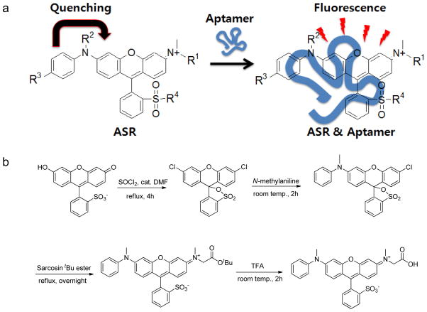
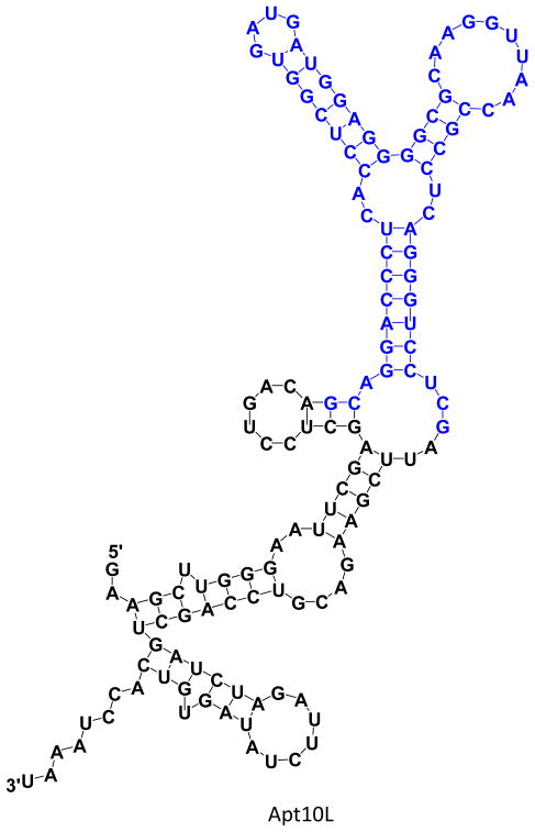
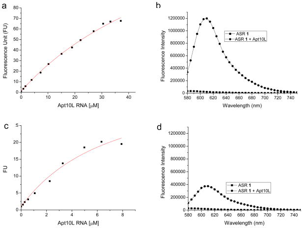
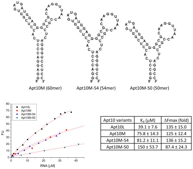
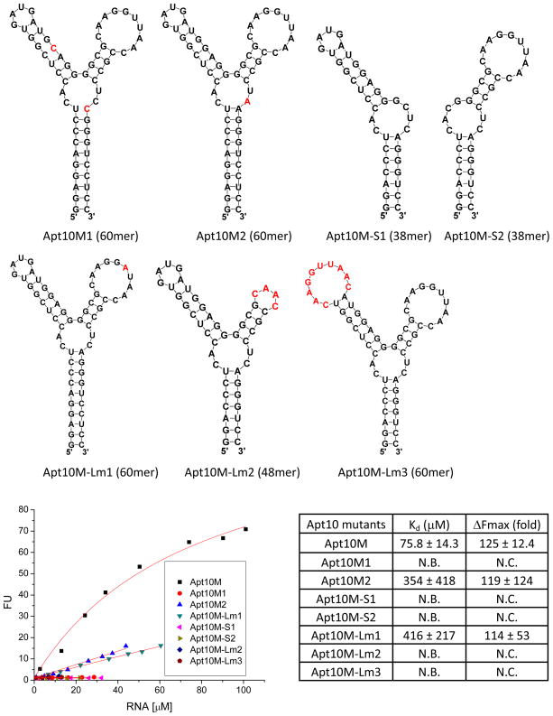
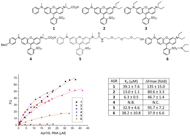
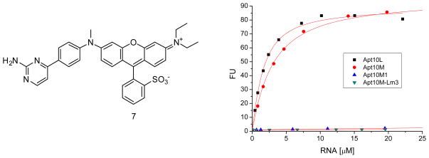
We report here a new small molecule fluorogen and RNA aptamer pair for RNA labeling. The small-molecule fluorogen is designed on the basis of fluorescently quenched sulforhodamine dye. The SELEX (Systematic Evolution of Ligands by EXponential enrichment) procedure and fluorescence screening in E. coli have been applied to discover the aptamer that can specifically activate the fluorogen with micromolar binding affinity. The systematic mutation and truncation study on the aptamer structure determined the minimum binding domain of the aptamer. A series of rationally modified fluorogen analogues have been made to probe the interacting groups of fluorogen with the aptamer. These results led to the design of a much improved fluorogen ASR 7 that displayed a 33-fold increase in the binding affinity for the selected aptamer in comparison to the original ASR 1 and an 88-fold increase in the fluorescence emission after the aptamer binding. This study demonstrates the value of combining in vitro SELEX and E. coli fluorescence screening with rational modifications in discovering and optimizing new fluorogen-RNA aptamer labeling pairs.


Figures

Figure 1
Figure 1
Design and synthesis of ASR fluorogenic probes for RNA tagging. a) The general structure of ASR and the scheme for the fluorescence enhancement of ASR by RNA aptamer binding (R1 = CH2CO2, R2 = Me, R3 = H, R4 = O for ASR 1 as the SELEX target). b) Synthetic scheme of the ASR 1 preparation.
Figure 2
Figure 2
Discovery of ASR binding aptamers via in vitro SELEX and E. coli fluorescence screening. a) Schematic of the selection strategy. b) Representative fluorescence image of a bacterial plate with 1 μM of ASR 1 in the LB Agar gel. Clones containing aptamers that enhance fluorescence of ASR 1 show increased fluorescence signal in the light-tight chamber of an IVIS 200 fluorescence imager: a colony with high fluorescence signal is marked with a red arrow.
Figure 3
Figure 3
Sequence and representative secondary structure of selected aptamer Apt10L as predicted by the M-fold program; total 4 structures are predicted for Apt10L. Sequences in blue represent the region of randomized 60 nucleotides. Apt10L contains long flanking sequences (HindIII site et al.) on 5′- and 3′-region for cloning into pBK-CMV-eGFP plasmid vector for fluorescence screening in E. coli.
Figure 4
Figure 4
Fluorescence titration of Apt10L RNA aptamer against ASR 1 (1 μM) in PBS buffer (pH 7.4) containing 1 mM MgCl2. Excitation and emission wavelength were 555 nm and 610 nm, respectively. a) The Kd value of Apt10L against ASR 1 is 39.1 ± 7.6 μM and Fmax (expected fluorescence enhancement at saturation) is 135 ± 15. Fluorescence intensity was normalized as fluorescence unit (FU) against the fluorescence intensity of ASR 1 in the absence of RNA. b) Fluorescence spectrum of ASR 1 in the absence or presence of several tens of μM Apt10L RNA. 610 nm is the maximum peak after RNA binding. c) and d) Fluorescence titrations were carried out using PAGE (6% polyarylamide-7M urea gel) purified Apt10L RNA at the same experimental conditions as in a) and b). The Kd value of Apt10L against ASR 1 is 3.5 ± 1.5 μM and Fmax is 29.0 ± 5.1.
Figure 5
Figure 5
Fluorescence titration of Apt10M variants against ASR 1 (1 μM) in PBS buffer (pH 7.4) containing 1 mM MgCl2. Excitation and emission wavelength were 555 nm and 610 nm, respectively.
Figure 6
Figure 6
Fluorescence titration of Apt10M mutants against ASR 1 (1 μM) in PBS buffer (pH 7.4) containing 1 mM MgCl2. Excitation and emission wavelength were 555 nm and 610 nm, respectively. N.C.: Not Calculable; N.B.: No Binding. Nucleotides in red stand for mutations.
Figure 7
Figure 7
Fluorescence titration of Apt10L against ASR analogues (1 μM) in PBS buffer (pH 7.4) containing 1 mM MgCl2. Excitation and emission wavelength were 555 nm and 610 nm, respectively. N.C.: Not calculable; N.B.: No binding.
Figure 8
Figure 8
ASR 7 containing a biaryl quencher has a highly improved binding affinity for Apt10L. Fluorescence titration was carried out in PBS buffer (pH 7.4) containing 1 mM MgCl2. Increasing concentrations of Apt10L RNA or selected Apt10M variants (Apt10M, Apt10M1 or Apt10M-Lm3) were added in the presence of 1 μM of ASR 7. Excitation and emission wavelength were 555 nm and 610 nm, respectively. Kd of ASR 7 for Apt10L RNA is 1.2 ± 0.1 μM and Fmax value is 88.6 ± 1.7. Kd of ASR 7 for Apt10M RNA is 2.6 ± 0.2 μM and Fmax value is 95.1 ± 1.5. Like ASR 1, ASR 7 does not bind Apt10M1 and Apt10M-Lm3 mutants, suggesting that the binding specificity not affected by the introduction of a biaryl quencher.

References

    1. Tsien RY. The green fluorescent protein. Annu Rev Biochem. 1998;67:509–544. - PubMed
    1. Marks KM, Nolan GP. Chemical labeling strategies for cell biology. Nat Methods. 2006;3:591–596. - PubMed
    1. Johnsson N, Johnsson K. Chemical tools for biomolecular imaging. ACS Chem Biol. 2007;2:31–38. - PubMed
    1. Fernandez-Suarez M, Ting AY. Fluorescent probes for super-resolution imaging in living cells. Nat Rev Mol Cell Biol. 2008;9:929–943. - PubMed
    1. Dragulescu-Andrasi A, Rao J. Chemical labeling of protein in living cells. ChemBioChem. 2007;8:1099–1101. - PubMed

Publication types