. 2010 Nov;27(11):1971-82.
doi: 10.1089/neu.2010.1470. Epub 2010 Oct 20.

Chondroitinase ABC enhances pericontusion axonal sprouting but does not confer robust improvements in behavioral recovery

Affiliations

Chondroitinase ABC enhances pericontusion axonal sprouting but does not confer robust improvements in behavioral recovery

Neil G Harris et al. J Neurotrauma. 2010 Nov.

Abstract

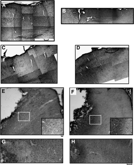
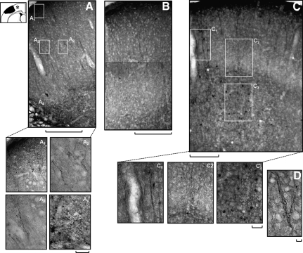
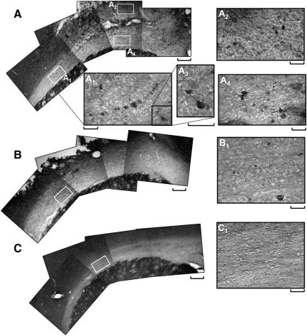
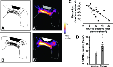
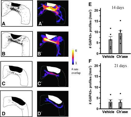
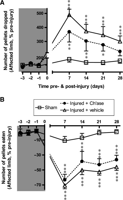
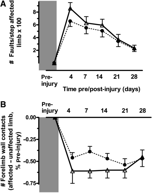
Traumatic brain injury (TBI) results in enduring functional deficits. Strategies aimed at promoting plasticity within the injured brain may aid in enhancing functional outcome. We have previously shown that spontaneous pericontusional axon sprouting occurs within 7-14 days after controlled cortical impact injury in the adult rat, but ultimately fails due to an increasingly growth-inhibitory environment. We therefore sought to determine whether acute infusion of chondroitinase ABC into the site of the cortical contusion, to further reduce pericontusional growth-inhibitory chondroitin sulfate proteoglycans (CSPGs), would enhance and prolong the sprouting response. We also wanted to determine if chondroitinase-enhanced sprouting would ameliorate the behavioral deficits in forelimb function that occur in this model. Acute chondroitinase infusion decreased intact CSPGs and significantly increased pericontusional cortical grey and white matter growth-associated protein 43 (GAP43)-positive axon sprouting at 7 days post-injury. A return of intact CSPGs at later time points likely contributed to the absence of persistently increased levels of axon sprouting by 14-21 days post-injury. There was no overall benefit on forelimb function during the time of maximal sprouting or at any subsequent times in three of four behavioral outcome measures. However, there was a chondroitinase-induced improvement in recovery from unskilled limb use deficits on the staircase forelimb reaching test toward sham-injured values at 28 days, which was not achieved by the vehicle-treated rats, indicating that there is some minor functional benefit of the increased sprouting induced by chondroitinase treatment. The current results, together with data from spinal cord injury models after chondroitinase intervention, suggest that a combinatorial approach with the addition of neurotrophins and rehabilitation would result in more robust axon sprouting and consequently improve behavioral outcome.


Figures

FIG. 1.
FIG. 1.
Representative sensorimotor cortical coronal immunohistochemistry brain sections at 7 days (AF) and 14 days (G and H) after injury, illustrating the degree of chondroitin sulfate proteoglycan (CSPG) digestion by chondroitinase in three different rats (A, B, C, E, and G), compared to vehicle-injected rats (D, F, and H). At 7 days after injury, sections stained with 2B6 primary antibody to visualize the CSPG “stubs” that remain after digestion revealed that in relatively intact tissue immediately anterior to the contusion, wide areas were affected by the enzyme (A), while tissue remaining in more severely-affected brains was visible along the pericontusional edge (B). In these same regions, staining for intact CSPGs showed a corresponding reduction in CSPGs in intact regions anterior to the contusion after chondroitinase (C), compared to vehicle-injected injured rats (D), and in more severely-affected rats after chondroitinase (E), compared to vehicle injection (F). Staining for intact CSPGs at 14 days after injury (G and H) revealed a developing glial scar in pericontusional tissue (left side of images) in both chondroitinase- (G) and vehicle-injected rats (H), together with similar levels of staining intensity and numerous CSPG-positive cells in both. This indicates reduced enzyme action and a return of inhibitory glycosylated CSPGs (scale bars = 250 μm for all images except insets [25 μm]).
FIG. 2.
FIG. 2.
Representative coronal immunohistochemistry brain sections of growth-associated protein 43 (GAP43) staining in grey matter sensorimotor cortex at 7 days after injury, illustrating enhanced axon sprouting after chondroitinase administration in two different brains at low power (A and C), and high power (A1A4, C1C3, and D), compared to a vehicle-treated, injured brain (B). No positive staining was ever noted in sham-injured rats (data not shown). The diagram at top left shows the approximate position of the stained regions (asterisk) relative to the injury site. Many of the stained grey matter fibers arose from GAP43-postive cell bodies in the deeper cortical layers (C and C3), and were often branched and appeared to extend dendrites (D). Other fibers followed a more tortuous route (A2), and showed bulbed endings (A3), especially close to the injury site (A4; scale bars = 250 μm in low-power images and 25 μm in high-power images).
FIG. 3.
FIG. 3.
Representative coronal immunohistochemistry brain sections of growth-associated protein 43 (GAP43) staining in the corpus callosum directly below the injury site at 7 days post-injury (A and B), or sham-injury (C), illustrating increased axon sprouting after chondroitinase treatment (A) compared to vehicle (B). Numerous GAP43-positive profiles were seen as black dots at low power after injury (A and B) compared to sham (C), and they were more prevalent after injury plus chondroitinase treatment (A) compared to injury plus vehicle (B). At high power, these positive profiles were observed as axonal fibers and as small bleb-like protuberances (A1A4 and B1) that were not present in sham-injured rats (C1; scale bars = 250 μm in low-power images and 25 μm in high-power images).
FIG. 4.
FIG. 4.
Co-registered semi-quantitative summed images constructed from six representative rats at the level of the bregma from post-injury day 7 injury plus vehicle (A and A′) and injury plus chondroitinase groups (B and B′), illustrating the difference in raw numbers of growth-associated protein 43 (GAP43)-positive profiles between groups (A and B), and the color-coded degree of overlap between the location of the positive cells among the six rats within each group (A′ and B′); see methods section for construction details. (C) Linear fit of a scatterplot showing the higher degree of correlation between injury severity (hemispheric loss of cortical volume) and the degree of axonal sprouting at 7 days after injury plus chondroitinase (solid squares, r = −0.71, p = 0.011, n = 10), compared to injury plus vehicle (open circles, r = −0.52, p = 0.15, n = 6). (D) Plot of numbers of GAP43-positive profiles in ipsilateral cortical grey and white matter, showing a significant increase in the number of profiles due to chondroitinase treatment (Ch'ase, n = 7) at 7 days after injury compared to vehicle (n = 4), after removing less-severely-injured rats (3 chondroitinase and 2 vehicle rats with <20% tissue loss, indicated above the dashed line in C) from both groups (*p < 0.05). Color image is available online at www.liebertonline.com/neu
FIG. 5.
FIG. 5.
Co-registered semi-quantitative summed images constructed from five representative rats at the level of the bregma from post-injury day 14 (A, B, A′, and B′) and day 21 (C, D, C′, and D′), from injury plus vehicle (A, A′, C, and C′), and injury plus chondroitinase groups (B, B′, D, and D′), illustrating the difference in raw numbers of growth-associated protein 43 (GAP43)-positive profiles between the groups at 14 days (A, B, C, and D) and the color-coded degree of overlap between the locations of the positive cells among the five rats within each group (A′, B′, C′, and D′). (E) Quantitative numbers of GAP43-profiles in ipsilateral grey and white matter at 14 days after injury. There was no significant difference at 14 days, although there was a trend towards increased sprouting due to chondroitinase treatment. (F) By 21 days there were reduced numbers of GAP43-positive profiles present in both groups, with no effect of treatment (Ch'ase, chondroitinase). Color image is available online at www.liebertonline.com/neu
FIG. 6.
FIG. 6.
Plots of unskilled (A) and skilled (B) affected forelimb function testing as evaluated by the staircase test in rats before and after injury plus chondroitinase treatment (Injured + Ch'ase, filled circles, n = 8), and injury plus vehicle (Injured + vehicle, open triangles), pooled vehicle treatment (n = 9), and injury only (n = 4) group after statistical testing), and in sham-injured rats (Sham, open squares, n = 9). Regardless of treatment, injury significantly increased the number of pellets retrieved and then dropped (unskilled limb use) at 7–21 days after injury (A), and decreased the number of pellets successfully retrieved and eaten at 7–28 days (B), compared to sham-injured rats (***p < 0.001). There was no significant difference between the two injury plus treatment groups when assessed by either parameter. However, unskilled use of the affected forelimb in the injury plus chondroitinase group was initially reduced relative to injury plus vehicle treatment, and improved considerably over time so that at 28 days the number of pellets dropped was not significantly different from the sham-injured group (**p < 0.01, *p < 0.05).
FIG. 7.
FIG. 7.
Tests of forelimb function using the grid-walk (A) and cylinder tests (B) in chondroitinase- (n = 9) and vehicle-treated injured rats (n = 14, solid circles and open triangles, respectively) in a separate cohort of rats from those used for the staircase test of forelimb reaching. Despite both tests yielding significant deficits in affected forelimb function out to 4 weeks post-injury when compared to the zero values seen pre-injury, indicating sustained sensitivity to injury, there was no significant benefit to chondroitinase administration seen at any time point post-injury. Similarly to the staircase test, there were minor trends in the data towards a positive effect of chondroitinase, particularly in the first week of treatment.

Similar articles

Cited by

References

    1. Asher R. Morgenstern D. Shearer M. Adcock K. Pesheva P. Fawcett J. Versican is upregulated in CNS injury and is a product of oligodendrocyte lineage cells. J. Neurosci. 2002;22:2225–2236. - PMC - PubMed
    1. Baker A.J. Phan N. Moulton R.J. Fehlings M.G. Yucel Y. Zhao M. Liu E. Tian G.F. Attenuation of the electrophysiological function of the corpus callosum after fluid percussion injury in the rat. J. Neurotrauma. 2002;19:587–599. - PubMed
    1. Bradbury E. Moon L. Popat R. King V. Bennett G. Patel P. Fawcett J. McMahon S. Chondroitinase ABC promotes functional recovery after spinal cord injury. Nature. 2002;416:636–640. - PubMed
    1. Bruckner G. Bringmann A. Hartig W. Koppe G. Delpech B. Brauer K. Acute and long-lasting changes in extracellular-matrix chondroitin-sulphate proteoglycans induced by injection of chondroitinase ABC in the adult rat brain. Exp. Brain Res. 1998;121:300–310. - PubMed
    1. Cafferty W.B. Yang S. Duffy P.J. Li S. Strittmatter S.M. Functional axonal regeneration through astrocytic scar genetically modified to digest chondroitin sulfate proteoglycans. J. Neurosci. 2007;27:2176–2185. - PMC - PubMed

Publication types

MeSH terms

Substances

LinkOut - more resources