Review
. 2011:73:29-46.
doi: 10.1146/annurev-physiol-012110-142145.

Heart valve structure and function in development and disease

Affiliations
Review

Heart valve structure and function in development and disease

Robert B Hinton et al. Annu Rev Physiol. 2011.

Abstract

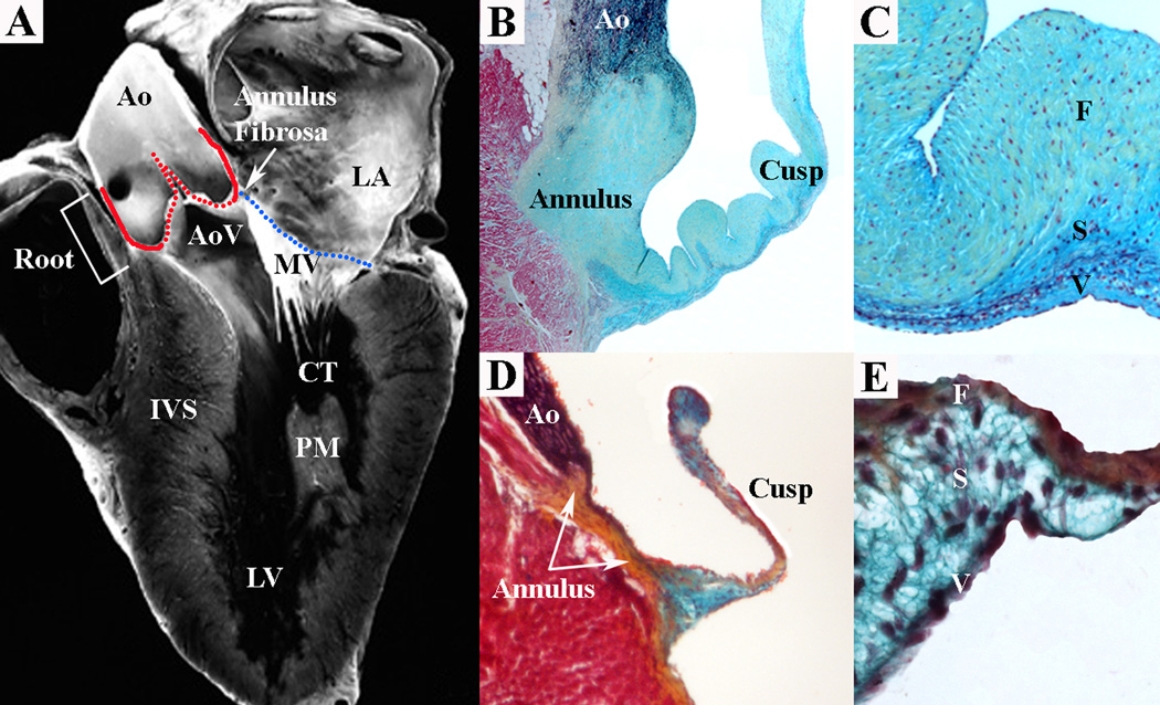
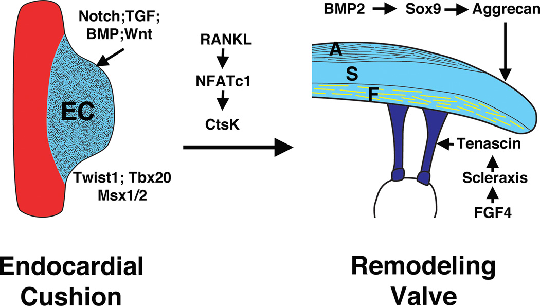
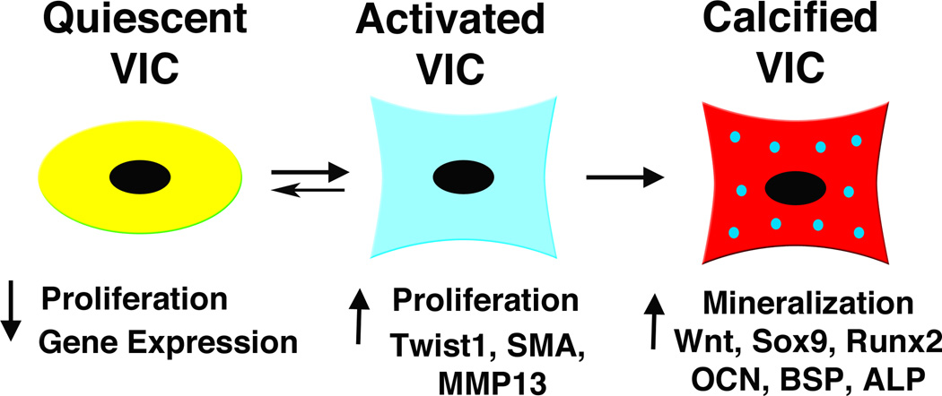
The mature heart valves are made up of highly organized extracellular matrix (ECM) and valve interstitial cells (VICs) surrounded by an endothelial cell layer. The ECM of the valves is stratified into elastin-, proteoglycan-, and collagen-rich layers that confer distinct biomechanical properties to the leaflets and supporting structures. Signaling pathways have critical functions in primary valvulogenesis as well as the maintenance of valve structure and function over time. Animal models provide powerful tools to study valve development and disease processes. Valve disease is a significant public health problem, and increasing evidence implicates aberrant developmental mechanisms underlying pathogenesis. Further studies are necessary to determine regulatory pathway interactions underlying valve pathogenesis in order to generate new avenues for novel therapeutics.


Figures

Figure 1
Figure 1
SL and AV valves with distinct structural and functional features are present in the human heart (A). The mitral valve (MV) is an AV valve and connects the left atrium (LA) to the left ventricle (LV). The MV consists of an annulus (A, blue line), leaflets and chordae tendineae (CT) that insert into papillary muscles (PM) in the myocardial wall. The aortic valve (AoV) is a SL valve and connects the LV to the aorta (Ao). The AoV consists of an annulus (A, red line) and cusps anchored within the aortic root (Root). Pentachrome staining shows valve ECM structure and composition in human (B,C) and mouse (D,E) aortic valves. At low magnification, SL valve tissue demonstrates cusp and annulus regions in human and mouse (B,D). At high magnification, aortic valve cusp architecture demonstrates similar ECM organization in human and mouse (C,E). The collagen-rich fibrosa layer (F) is oriented on the arterial aspect of the cusp, while the elastin-rich ventricularis layer (V) is oriented on the ventricular aspect of the cusp. The proteoglycan-rich spongiosa layer (S) interconnects the collagen and elastin fibers. IVS interventricular septum. (Panel A from reference (115), with permission.)
Figure 2
Figure 2
Regulatory interactions of signaling pathways and transcription factors in heart valve development. Signaling pathways including Notch, Transforming growth factor (TGF), Bone morphogenetic protein (BMP), and Wnt, with transcription factors including Twist1, Tbx20 and Msx1/2 are involved in endocardial cushion (EC) formation during early valvulogenesis. NFATc1 signaling contributes to elongation and remodeling of the EC. During valve maturation, BMP signaling induces cartilage-associated genes Sox9 and aggrecan. Fibroblast growth factor (FGF) signaling promotes expression of scleraxis and tenascin, which are characteristic of tendon cell lineages. These genes and pathways involved in valve development also are active in adult valve disease.
Figure 3
Figure 3
Valve interstitial cell (VIC) phenotype relates to maladaptive and pathologic signaling pathways. Quiescent VICs show little proliferation or gene expression, while activated VICs demonstrate increased proliferation and increased myofibroblast-associated gene expression. VIC activation may be adaptive or maladaptive, and patterns of signaling pathway gene expression may distinguish these features. Some maladaptive VIC activation and induction of genes associated with bone formation are apparent in valve tissue calcification. SMA smooth muscle α-actin; MMP matrix metalloprotease; OCN osteocalcin; BSP bone sialoprotein; ALP alkaline phosphatase.

References

    1. Schoen FJ. Evolving concepts of cardiac valve dynamics. Circulation. 2008;118:1864–1880. - PubMed
    1. Armstrong EJ, Bischoff J. Heart valve development: Endothelial cell signaling and differentiation. Circ Res. 2004;95:459–470. - PMC - PubMed
    1. Bruneau BG. The developmental genetics of congenital heart disease. Nature. 2008;451:943–948. - PubMed
    1. Sacks MS, David Merryman W, Schmidt DE. On the biomechanics of heart valve function. J Biomech. 2009;42:1804–1824. - PMC - PubMed
    1. Hoffman JIE, Kaplan S. The incidence of congenital heart disease. J Am Coll Cardiol. 2002;39:1890–1900. - PubMed

Publication types