. 2010 Sep 1:5:22.
doi: 10.1186/1749-8104-5-22.

Meis1 specifies positional information in the retina and tectum to organize the zebrafish visual system

Affiliations

Meis1 specifies positional information in the retina and tectum to organize the zebrafish visual system

Timothy Erickson et al. Neural Dev. .

Abstract

Background: During visual system development, multiple signalling pathways cooperate to specify axial polarity within the retina and optic tectum. This information is required for the topographic mapping of retinal ganglion cell axons on the tectum. Meis1 is a TALE-class homeodomain transcription factor known to specify anterior-posterior identity in the hindbrain, but its role in visual system patterning has not been investigated.

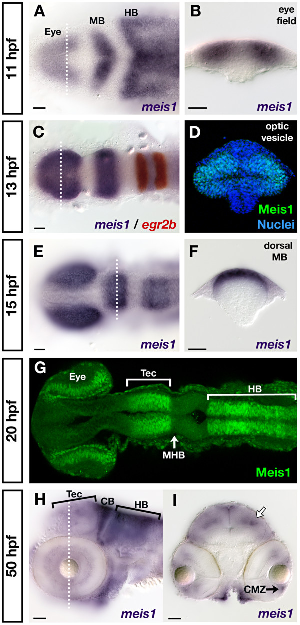
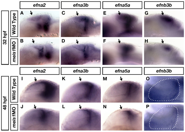
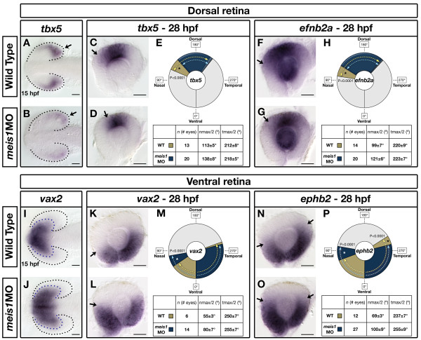
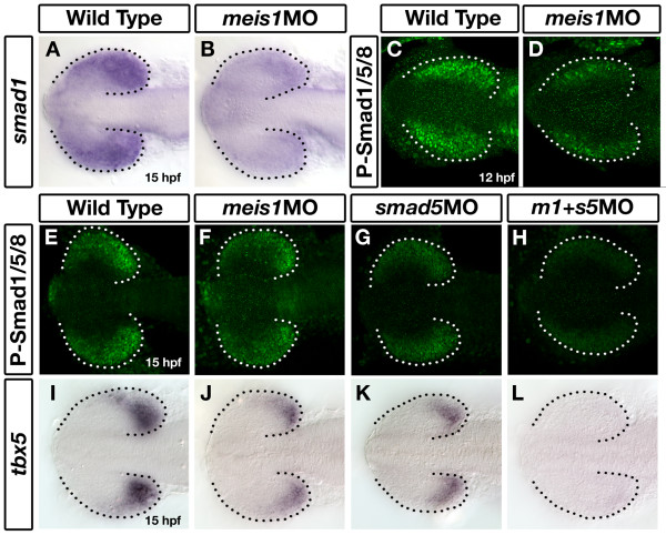
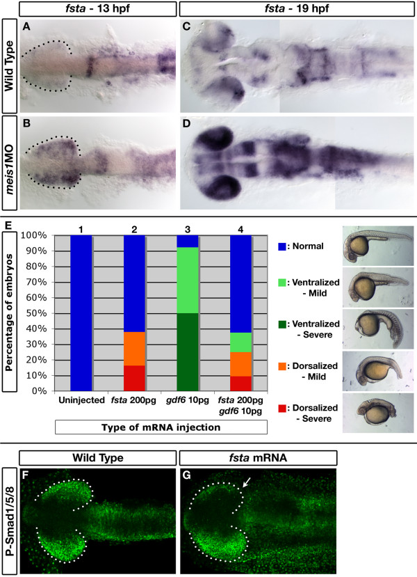
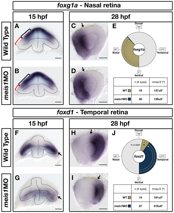
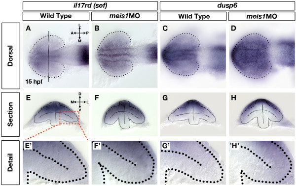
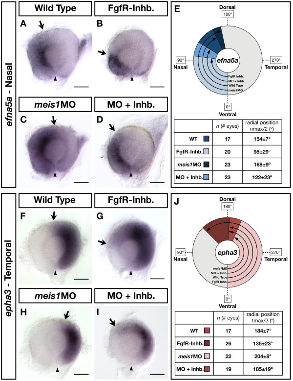
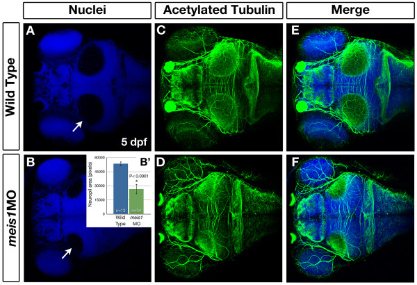
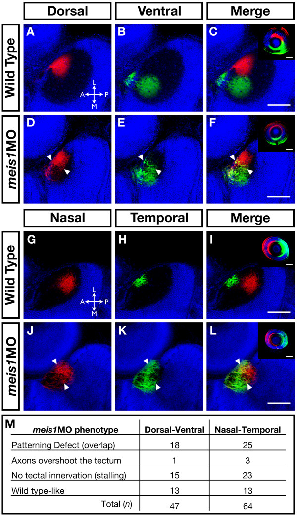
Results: meis1 is expressed in both the presumptive retina and tectum. An analysis of retinal patterning reveals that Meis1 is required to correctly specify both dorsal-ventral and nasal-temporal identity in the zebrafish retina. Meis1-knockdown results in a loss of smad1 expression and an upregulation in follistatin expression, thereby causing lower levels of Bmp signalling and a partial ventralization of the retina. Additionally, Meis1-deficient embryos exhibit ectopic Fgf signalling in the developing retina and a corresponding loss of temporal identity. Meis1 also positively regulates ephrin gene expression in the tectum. Consistent with these patterning phenotypes, a knockdown of Meis1 ultimately results in retinotectal mapping defects.

Conclusions: In this work we describe a novel role for Meis1 in regulating Bmp signalling and in specifying temporal identity in the retina. By patterning both the retina and tectum, Meis1 plays an important role in establishing the retinotectal map and organizing the visual system.


Figures

Figure 1
Figure 1
Developmental time course of meis1 mRNA and protein expression. (A, B) mRNA in situ hybridizations (ISHs) for meis1 at 11 hpf showing expression in the eye field, and in the presumptive midbrain (MB) and hindbrain (HB). The transverse section (B) shows meis1 expression in the eye field. (C) mRNA ISH showing meis1 expression at 13 hpf in the optic vesicles, midbrain and hindbrain. egr2b/krox20 expression (red) marks rhombomeres 3 and 5 of the hindbrain. (D) Transverse section of a whole mount immunostain for Meis1 showing protein in the dorsal and ventral leaflets of the 13 hpf optic vesicle. Hoechst 33258 stain marks the nuclei. (E, F) mRNA ISH at 15 hpf showing continued expression of meis1 in the optic vesicles, midbrain and hindbrain. The transverse section (F) shows meis1 expression in the dorsal midbrain. (G) Whole mount immunostain for Meis1 protein at 20 hpf. Meis1 protein is present in the eye, presumptive tectum (Tec), and in the hindbrain (HB) up to the r1-r2 boundary. Meis1 is excluded from the midbrain-hindbrain boundary (MHB). (H, I) mRNA ISH at 50 hpf showing meis1 expression in the hindbrain (HB) and cerebellum (CB) and tectum (Tec). The transverse section (I) shows meis1 expression in the ciliary marginal zone (CMZ) of the retina and in the dorsal midline and a deeper layer of the tectum (white arrow). Embryos in (A, C, E, G) are shown in dorsal view with anterior to the left. Embryo in (H) is shown in lateral view with anterior to the left. Transverse sections in (B, D, F, I) are oriented dorsal up. The dotted lines in (A, C, E, H) indicate the position of the corresponding transverse sections in (B, D, F, I). All scale bars = 50 μm.
Figure 2
Figure 2
Meis1 positively regulates ephrin gene expression in the tectum. (A-H) mRNA in situ hybridizations (ISHs) for efna2 (A, B), efna3b (C, D), efna5a (E, F) and efnb3b (G, H) in 32-hpf wild-type (A, C, E, G) and meis1 morphant (meis1MO) (B, D, F, H) embryos. Meis1 knockdown leads to a downregulation in the tectal expression of these ephrin genes (arrows) (I-P) mRNA ISH for efna2 (I, J), efna3b (K, L), efna5a (M, N) and efnb3b (O, P) in 48-hpf wild-type (I, K, M, O) and meis1 morphant (J, L, N, P) embryos. The early defects in tectal ephrin gene expression remain in 48-hpf meis1 morphants (arrows). The dotted lines in (O, P) outline a single tectal lobe in each embryo. Embryos in (A-N) are shown in lateral view with anterior left, while embryos in (O, P) are shown in dorsal view with anterior left.
Figure 3
Figure 3
Meis1 contributes to dorsal-ventral patterning in the developing retina. (A, B, I, J) mRNA in situ hybridization (ISH) for the dorsal marker tbx5 (A, B) and the ventral marker vax2 (I, J) in 15-hpf wild-type and meis1 morphant (meis1MO) embryos. Black dotted lines outline the optic vesicle. Arrows indicate tbx5 expression in the presumptive dorsal retina. Purple dotted lines indicate the domain of vax2 expression. All views are dorsal with anterior left. (C, D, F, G, K, L, N, O) mRNA ISH for dorsal genes tbx5 (C, D) and efnb2a (F, G), and ventral genes vax2 (K, L) and ephb2 (N, O) in dissected, flat-mounted eyes from 28-hpf wild-type and meis1 morphant embryos. Arrows indicate the approximate limit of the gene expression domain. (E, H, M, P) The domains of gene expression were quantified by determining a 360° profile of in situ staining intensity and graphing the radial position at which gene expression intensity falls to the halfway point between its minimum and maximum values. The nmax/2 and tmax/2 values are given as the mean radial position in degrees ± one standard deviation. Asterisks indicate regions in which there are statistically significant differences in axial identity between wild type (WT) and meis1 morphants as determined by an unpaired, two-tailed t-test using a P-value of 0.01 as a cutoff for significance. Representative dissected eyes are shown. Scale bars = 50 μm.
Figure 4
Figure 4
Meis1 positively regulates smad1 expression in the developing eye. (A, B) mRNA in situ hybridization (ISH) for smad1 in 15-hpf wild-type and meis1 morphant (meis1MO) embryos. (C, D) Whole mount immunostains for phosphorylated Smad1/5/8 on 12-hpf wild-type and meis1 morphant embryos. (E-H) Phospho-Smad1/5/8 immunostains on 15-hpf embryos treated with meis1 morpholino (F), smad5 morpholino (G) or a combination of both morpholinos (H). (I-L) mRNA ISH for tbx5 in 15-hpf wild-type (I), meis1 morphant (J), smad5 morphant (K), and meis1-smad5 double morphant (L) embryos. Dotted lines outline the optic vesicle. All views are dorsal with anterior to the left.
Figure 5
Figure 5
follistatin a is ectopically expressed in Meis1-depleted embryos and can inhibit Gdf6-mediated Bmp signalling. (A-D) mRNA in situ hybridizations for fsta on 13-hpf (A, B) and 19-hpf (C, D) wild-type and meis1 morphant (meis1MO) embryos. Dotted lines outline the optic vesicle. All views are dorsal with anterior to the left. (E) Results of the GDF6-Fsta interaction experiments. One-cell embryos were injected with either 200 pg of zebrafish fsta mRNA (bar 2), human GDF6 mRNA (bar 3), or both mRNAs (bar 4), raised until 28 hpf, and scored for dorsalized and ventralized phenotypes (see legend on the right for classification). (F, G) Confocal images of whole mount immunostains for phospho-Smad1/5/8 in wild-type and fsta mRNA-injected embryos at 14 hpf. Injection of fsta mRNA into one cell of a two-cell embryo causes a unilateral reduction in phospho-Smad1/5/8 staining (arrow in G). Dotted lines outline the optic vesicle. Views are dorsal with anterior to the left.
Figure 6
Figure 6
Meis1-depleted embryos exhibit a partial temporal-to-nasal shift in retinal identity. (A, B, F, G) mRNA in situ hybridization (ISH) for the nasal marker foxg1a (A, B) and the temporal marker foxd1 (F, G) in 15-hpf wild-type and meis1 morphant (meis1MO) embryos. Dotted lines outline the optic vesicle. The black brackets in (A, B) indicate the proximal-distal extent of foxg1a expression, while the red brackets indicate the foxg1a-free region. Arrows in (F, G) indicate the dorsal leaflet of the optic vesicle. Transverse sections are oriented dorsal up. (C, D, H, I) mRNA ISH for the nasal marker foxg1a (C, D) and the temporal marker foxd1 (H, I) in dissected, flat-mounted eyes from 28-hpf wild-type and meis1 morphant embryos. The arrows indicate the approximate limit of the gene expression domain. (E, J) The domains of gene expression were quantified by determining a 360° profile of in situ staining intensity and graphing the radial position at which gene expression intensity falls to the halfway point between its minimum and maximum values. The nmax/2 and tmax/2 values are given as the mean radial position in degrees ± one standard deviation. Asterisks indicate regions in which there are statistically significant differences in axial identity between wild type and meis1 morphants as determined by an unpaired, two-tailed t-test using a P-value of 0.01 as a cutoff for significance. Representative dissected eyes are shown. Scale bars = 50 μm.
Figure 7
Figure 7
Retinal Fgf signalling is upregulated in Meis1-depleted embryos. (A-D) mRNA in situ hybridization (ISH) for the Fgf-responsive genes il17rd/sef (A, B) and dusp6 (C, D) in wild-type (A, C) and meis1 morphant (meis1MO) (B, D) embryos. Dotted lines outline the optic vesicle. The vertical dotted line in (A) indicates the estimated position of the transverse sections in (E-H). Views are dorsal with anterior left. (E-H) Transverse sections through the eyes of 15-hpf wild-type and meis1 morphant embryos stained for il17rd and dusp6. (E'-H') Detailed views of the corresponding sections in (E-H). The region of interest is indicated by the red dashed-line box. Dotted lines outline the optic vesicles. All transverse sections are oriented with dorsal up. Legend for retinal axial position: D, dorsal; V, ventral; N, nasal; T, temporal; L, lateral; M, medial; A, anterior; P, posterior.
Figure 8
Figure 8
The contribution of Fgf signalling to the NT patterning defects in Meis1-depleted embryos. (A-D, F-I) mRNA in situ hybridizations for the NT markers efna5a (A-D) and epha3 (F-I) in wild type, Meis1-depleted (meis1MO), Fgf receptor-inhibitor treated (FgfR-Inhb.), and FgfR-inhibited/Meis1-depleted retinas (MO + Inhb.). Arrows indicate the extent of the gene expression domain, while the arrowheads indicate the position of the ventral choroid fissure. Representative dissected eyes are shown oriented with dorsal up and nasal to the left. Scale bars = 50 μm. (E, J) Quantification of the changes in efna5a and epha3 expression, as quantified by measuring a 360° profile of in situ staining intensity and graphing the mean radial position at which gene expression intensity falls to the halfway point between its minimum and maximum values. The nmax/2 and tmax/2 values are given as the mean radial position in degrees ± one standard deviation. WT, wild type.
Figure 9
Figure 9
Meis1-depleted embryos have smaller tectal neuropil at 5 dpf. (A-F) Whole mount immunohistochemistry using anti-acetylated tubulin (axons - green) and Hoechst 33258 (nuclei - blue) to compare the size of the tectal neuropil in 5-dpf wild type (A, C, E) and meis1 morphant (meis1MO) (B, D, F) embryos. White arrows in (A, B) indicate the tectal neuropil. (B') The area (in pixels) of the neuropil from wild type and meis1 morphant embryos was measured using ImageJ. The n values represent individual neuropil regions. The error bars show plus/minus one standard deviation. The asterisk indicates a statistically significant reduction the size of morphant neuropil as determined by an unpaired, two-tailed t-test.
Figure 10
Figure 10
The retinotectal map is disorganized in Meis1-depleted embryos. (A-L) Lipophilic fluorescent dyes DiI (red) and DiO (green) were injected into specific axial positions of the retina of fixed 5-dpf wild-type and meis1 morphant embryos and innervation patterns of the ganglion cell axons on the tectum were imaged by confocal microscopy. Nuclei are stained with Hoechst 33258 (blue). The wild-type (A-C) medial-lateral segregation of dorsal (red) and ventral (green) ganglion cell axons in the tectum is lost in meis1 morphants (white arrowheads in (D-F)). Similarly, the wild-type (G-I) anterior-posterior segregation of nasal (red) and temporal (green) ganglion cell axons in the tectum is disorganized in meis1 morphants (white arrowheads in (J-L)). The insets in (C, F, I, L) are lateral views of injected retinas from the embryos shown in the corresponding panels. Retinas are oriented with dorsal up and nasal to the left, while all tectal views are dorsal with anterior to the left. Legend for axial position in the tectum: L, lateral; M, medial; A, anterior; P, posterior. All scale bars = 75 μm. (M) Table describing the frequency of various retinotectal mapping phenotypes observed in meis1 morphants (meis1MO). No retinotectal mapping defects were observed in any wild-type embryos examined.

Similar articles

Cited by

References

    1. Scicolone G, Ortalli AL, Carri NG. Key roles of Ephs and ephrins in retinotectal topographic map formation. Brain Res Bull. 2009;79:227–247. doi: 10.1016/j.brainresbull.2009.03.008. - DOI - PubMed
    1. Lemke G, Reber M. Retinotectal mapping: new insights from molecular genetics. Annu Rev Cell Dev Biol. 2005;21:551–580. doi: 10.1146/annurev.cellbio.20.022403.093702. - DOI - PubMed
    1. McLaughlin T, Hindges R, O'Leary DD. Regulation of axial patterning of the retina and its topographic mapping in the brain. Curr Opin Neurobiol. 2003;13:57–69. doi: 10.1016/S0959-4388(03)00014-X. - DOI - PubMed
    1. Harada T, Harada C, Parada LF. Molecular regulation of visual system development: more than meets the eye. Genes Dev. 2007;21:367–378. doi: 10.1101/gad.1504307. - DOI - PubMed
    1. Gosse NJ, Baier H. An essential role for Radar (Gdf6a) in inducing dorsal fate in the zebrafish retina. Proc Natl Acad Sci USA. 2009;106:2236–2241. doi: 10.1073/pnas.0803202106. - DOI - PMC - PubMed

Publication types

MeSH terms