. 2010 Sep 1:3:17.
doi: 10.1186/1755-1536-3-17.

Mast cells and hypoxia drive tissue metaplasia and heterotopic ossification in idiopathic arthrofibrosis after total knee arthroplasty

Affiliations

Mast cells and hypoxia drive tissue metaplasia and heterotopic ossification in idiopathic arthrofibrosis after total knee arthroplasty

Theresa A Freeman et al. Fibrogenesis Tissue Repair. .

Abstract

Background: Idiopathic arthrofibrosis occurs in 3-4% of patients who undergo total knee arthroplasty (TKA). However, little is known about the cellular or molecular changes involved in the onset or progression of this condition. To classify the histomorphologic changes and evaluate potential contributing factors, periarticular tissues from the knees of patients with arthrofibrosis were analyzed for fibroblast and mast cell proliferation, heterotopic ossification, cellular apoptosis, hypoxia and oxidative stress.

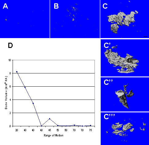
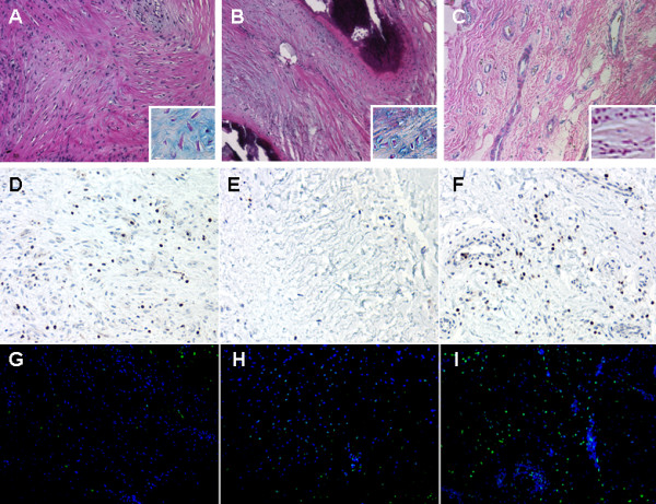
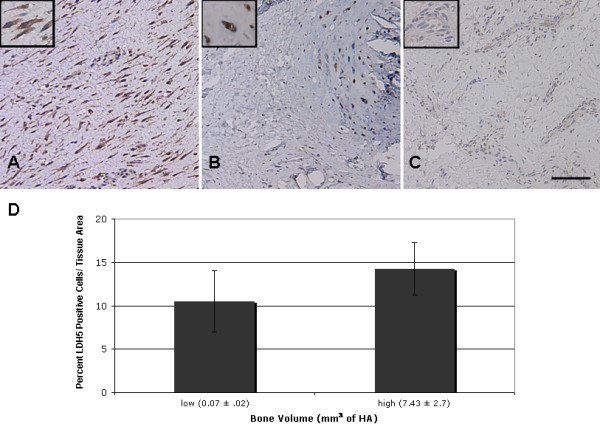
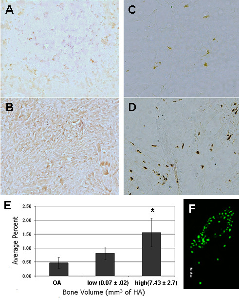
Results: The arthrofibrotic tissue was composed of dense fibroblastic regions, with limited vascularity along the outer edges. Within the fibrotic regions, elevated numbers of chymase/fibroblast growth factor (FGF)-expressing mast cells were observed. In addition, this region contained fibrocartilage and associated heterotopic ossification, which quantitatively correlated with decreased range of motion (stiffness). Fibrotic, fibrocartilage and ossified regions contained few terminal dUTP nick end labeling (TUNEL)-positive or apoptotic cells, despite positive immunostaining for lactate dehydrogenase (LDH)5, a marker of hypoxia, and nitrotyrosine, a marker for protein nitrosylation. LDH5 and nitrotyrosine were found in the same tissue areas, indicating that hypoxic areas within the tissue were associated with increased production of reactive oxygen and nitrogen species.

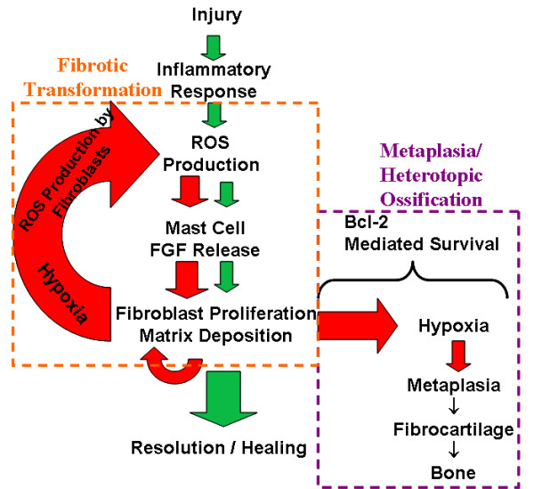
Conclusions: Taken together, we suggest that hypoxia-associated oxidative stress initiates mast cell proliferation and FGF secretion, spurring fibroblast proliferation and tissue fibrosis. Fibroblasts within this hypoxic environment undergo metaplastic transformation to fibrocartilage, followed by heterotopic ossification, resulting in increased joint stiffness. Thus, hypoxia and associated oxidative stress are potential therapeutic targets for fibrosis and metaplastic progression of idiopathic arthrofibrosis after TKA.


Figures

Figure 1
Figure 1
Microcomputed tomography (microCT) analysis of the periarticular tissue and comparison of bone volume (BV) and range of motion (ROM). Representative microCT images are shown for (A) minimal calcification in pre-surgical osteoarthritic tissues (n = 5), (B) low BV group (0.07 ± 0.02 mm3 of hydroxyapatite; n = 5) and (C) high BV group (7.4 ± 2.7 mm3; n = 5). Calcified regions adjoining the fibrocartilage were confirmed by microCT analysis to have the appearance and density consistent with newly formed bone. (D) ROM and BV comparisons; the closer the patient ROM was to normal flexion, the lower the amount of tissue calcification or BV. Once the ROM was restricted to < 50 degrees, BV increased, and reached a peak when ROM was ≤ 20 degrees.
Figure 2
Figure 2
Tissue morphology, cell survival and programmed cell death in periarticular tissue. Representative images are shown for (A, D, G) fibrotic (B, E, H), fibrocartilage with calcification (C, F, I) and vascular regions. Tissues were stained (A-C)with hematoxylin and eosin and alcian blue (insets enlarged 100%), (D-F) Bcl-2 and terminal dUTP nick end labeling (TUNEL); (G-I) green apoptotic cells and 4',6-diamidino-2-phenylindole(DAPI)-stained blue stained nuclei. Scale bar = 100 μm.
Figure 3
Figure 3
Immunohistochemical stain of arthrofibrotic tissue for lactate dehydrogenase (LDH)5. Representative LDH5 immunohistochemical results are shown for (A) fibrotic regions, (B) fibrocartilage/calcified regions and (C) vascular regions of the tissue. (D) Image analysis of LDH5 for the low and high bone volume (BV) groups. There was no significant difference between the two groups. The high magnification insets emphasize the increased LDH5 expression in fibroblasts and chondrocytes. Scale bar = 250 μm.
Figure 4
Figure 4
Immunohistochemical stain of arthrofibrotic tissue for nitrotyrosine, chymase and fibroblast growth factor (FGF). Representative immunohistochemical results are shown for nitrotyrosine in (A) osteoarthritis (OA) tissue and (B) arthrofibrotic tissue, and chymase in (C) OA tissue and (D) arthrofibrotic tissue. (E) Image analysis of mast cell numbers for the low and high bone volume (BV) groups, compared with the OA cohort. There was a significant difference between all three groups (P < 0.05). (F) Representative immunohistochemical result of FGF expression in mast cells.
Figure 5
Figure 5
Summary of findings of arthrofibrotic progression. Green arrows indicate normal healing pathway, red arrows highlight disease progression pathway. The orange box indicates the etiology of the oxidative fibrotic transformation process supported by our previous studies [13]. The purple box indicates the pathology of metaplastic heterotopic ossification, supported by this study.

Similar articles

Cited by

References

    1. Anouchi YS, McShane M, Kelly F, Elting J, Stiehl J. Range of motion in total knee replacement. Clinical Orthopaedics & Related Research. 1996;331:87–92. - PubMed
    1. Markel DC, Luessenhop CP, Windsor RE, Sculco TA. Arthroscopic treatment of peripatellar fibrosis after total knee arthroplasty. Journal of Arthroplasty. 1996;11:293–297. doi: 10.1016/S0883-5403(96)80081-7. - DOI - PubMed
    1. Kim J, Nelson CL, Lotke PA. Stiffness after total knee arthroplasty. Prevalence of the complication and outcomes of revision. Journal of Bone & Joint Surgery American. 2004;86:1479–1484. - PubMed
    1. Mont MA, Serna FK, Krackow KA, Hungerford DS. Exploration of radiographically normal total knee replacements for unexplained pain. Clinical Orthopaedics & Related Research. 1996;331:216–220. - PubMed
    1. Nicholls DW, Dorr LD. Revision surgery for stiff total knee arthroplasty. Journal of Arthroplasty. 1990;5(Suppl):S73–7. doi: 10.1016/S0883-5403(08)80029-0. - DOI - PubMed

LinkOut - more resources