. 2010 Sep 1:5:23.
doi: 10.1186/1749-8104-5-23.

Migration, early axonogenesis, and Reelin-dependent layer-forming behavior of early/posterior-born Purkinje cells in the developing mouse lateral cerebellum

Affiliations

Migration, early axonogenesis, and Reelin-dependent layer-forming behavior of early/posterior-born Purkinje cells in the developing mouse lateral cerebellum

Takaki Miyata et al. Neural Dev. .

Abstract

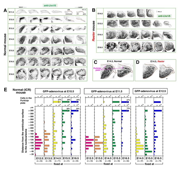
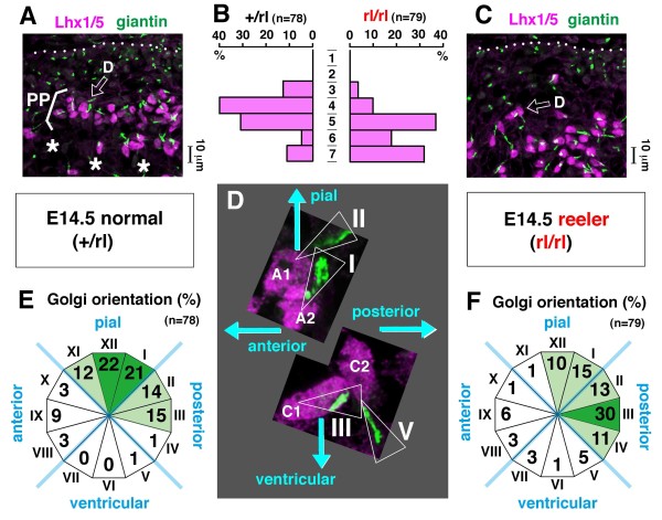
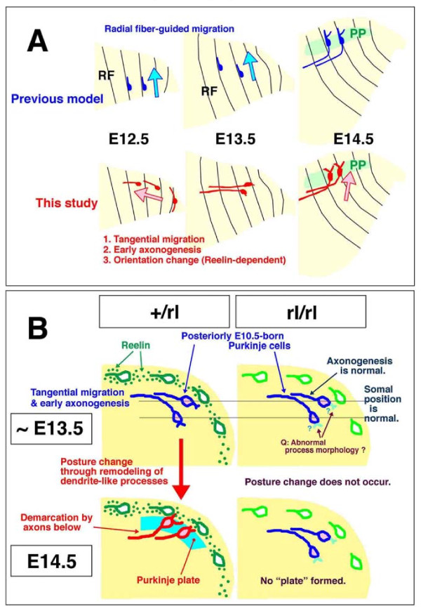
Background: Cerebellar corticogenesis begins with the assembly of Purkinje cells into the Purkinje plate (PP) by embryonic day 14.5 (E14.5) in mice. Although the dependence of PP formation on the secreted protein Reelin is well known and a prevailing model suggests that Purkinje cells migrate along the 'radial glial' fibers connecting the ventricular and pial surfaces, it is not clear how Purkinje cells behave in response to Reelin to initiate the PP. Furthermore, it is not known what nascent Purkinje cells look like in vivo. When and how Purkinje cells start axonogenesis must also be elucidated.

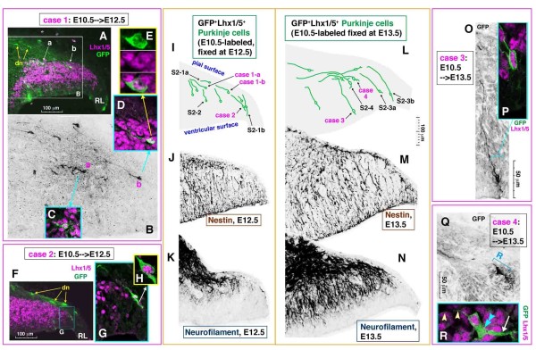
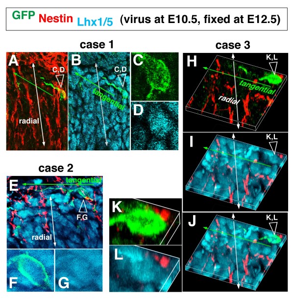
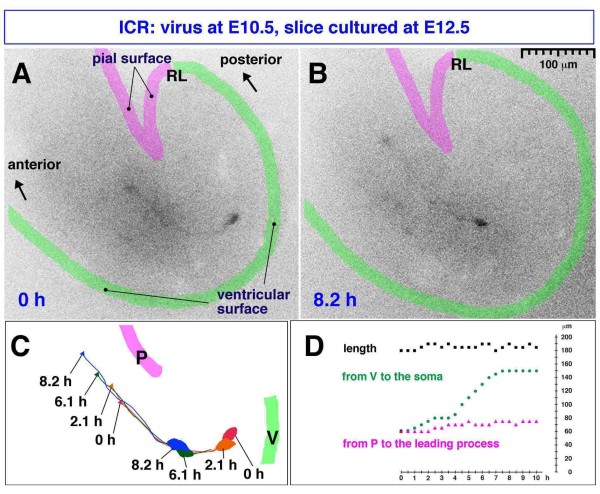
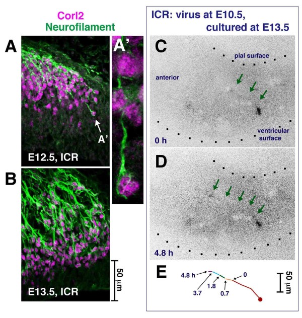
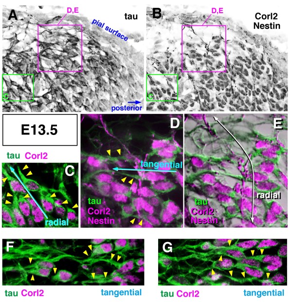
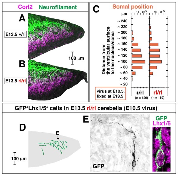
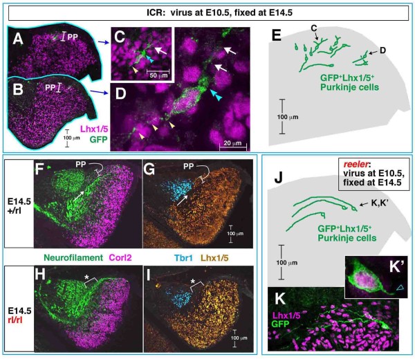
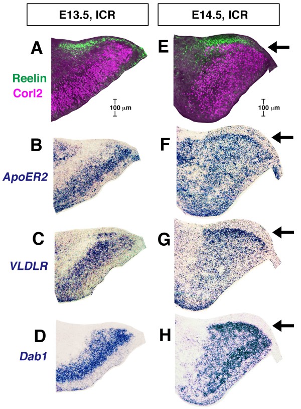
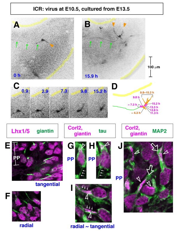
Results: We show that Purkinje cells generated on E10.5 in the posterior periventricular region of the lateral cerebellum migrate tangentially, after only transiently migrating radially, towards the anterior, exhibiting an elongated morphology consistent with axonogenesis at E12.5. After their somata reach the outer/dorsal region by E13.5, they change 'posture' by E14.5 through remodeling of non-axon (dendrite-like) processes and a switchback-like mode of somal movement towards a superficial Reelin-rich zone, while their axon-like fibers remain relatively deep, which demarcates the somata-packed portion as a plate. In reeler cerebella, the early born posterior lateral Purkinje cells are initially normal during migration with anteriorly extended axon-like fibers until E13.5, but then fail to form the PP due to lack of the posture-change step.

Conclusions: Previously unknown behaviors are revealed for a subset of Purkinje cells born early in the posteior lateral cerebellum: tangential migration; early axonogenesis; and Reelin-dependent reorientation initiating PP formation. This study provides a solid basis for further elucidation of Reelin's function and the mechanisms underlying the cerebellar corticogenesis, and will contribute to the understanding of how polarization of individual cells drives overall brain morphogenesis.


Figures

Figure 1
Figure 1
Stage-dependent changes of the overall distribution of Purkinje cells in embryonic cerebella demonstrated with Lhx1/5 immunohistochemistry and adenovirus-mediated pulse-chase methods. (A-D) Anti-Lhx1/5 immunostained sagittal sections of normal (ICR or +/rl) (A, C) and reeler (rl/rl) (B, D) cerebella. Sections in each row of (A, B) are arranged in a lateral-to-medial order. The dorsal side is on the top of each picture and the posterior side is on the right. At E14.5, the Purkinje plate (PP) emerges in the normal (C) but not in reeler (D) cerebellum, and this initial difference became progressively enhanced until the perinatal period (E18.5), when the subpial layering versus deeply clustered patterns became complete. (E) Histogram showing the distance of green fluorescent protein (GFP) and Lhx1/5 double positive cells from the ventricular surface at the indicated stages (bottom) following Lyn-Venus adenovirus injections at E10.5, E11.5, and E12.5. Consistent with a general tendency that cells born earlier migrate further, the cells that contributed best to the emergence of the PP at E14.5 were born on E10.5 (presented separately at the top of the graph). Note, however, that some of the E10.5-born cells are seen deeply (within 150 μm from the ventricular surface) even at E15.5 and E16.5.
Figure 2
Figure 2
The early/posterior-born Purkinje cells are elongated radially and tangentially in the lateral cerebellum at E12.5 and E13.5. (A-Q) The morphology and orientation of the E10.5-adenovirally labeled nascent Purkinje cells in E12.5 (A-I) and E13.5 (L, O, Q) cerebella are compared with immunoreactivity for Nestin (J, M) (almost identical staining patterns were obtained by using RC2 (not shown)) and Neurofilament (K, N). dn, prospective deep nuclear neurons; RL, rhombic lip. In (I, L), traces of representative cases of E10.5-born nascent Purkinje cells (including cases 1a, 1b, 2, 3, and 4 in this figure, as well as those shown in Additional file 2) are summarized in an illustration of a 'standardized' cerebellar primordium at E12.5 or E13.5. The scale is common in panels (I-N). Note that E10.5-born Purkinje cells seen in an outer zone near the pial surface are tangentially elongated whereas those in deeper regions are more radially elongated. Neurofilament+ radial fibers markedly increased between E12.5 and E13.5 and appear to outnumber Nestin+ fibers at E13.5. (R) Each tangentially oriented Purkinje cell at E13.5 (Q, R) is polarized with a single thin and long process extended anteriorly (arrowheads) and a thick cytoplasmic part (double blue arrowhead) from which a few thick processes (arrow) are extended posteriorly.
Figure 3
Figure 3
Triple immunolabeling analysis to demonstrate the relationship between tangentially oriented nascent Purkinje cells and radial fibers. (A-G, K, L) Sections as used for the E10.5 to E12.5 tracing analysis (Figure 2A-I) were stained with anti-GFP (green), anti-Lhx1/5 (cyan), and anti-Nestin (red). In each of the three representative cases, magnified views for the somal part (open arrowhead) are provided to confirm Lhx1/5 immunoreacivity. (H-L) In case 3, three-dimensional reconstructions are shown.
Figure 4
Figure 4
Time-lapse monitored tangential migration of a Purkinje-like cell. (A, B) A Venus-expressing E10.5-born Purkinje-like cell in an E12.5 cerebellar slice (see also Additional file 3). (C) Traces of migration: P, pial surface; V, ventricular surface; arrow, tip of the leading process. (D) Graph depicting the length of the observed cell, distance from the ventricular surface to the soma, and distance from the pial surface to the tip of leading process. Migration of this cell is characterized by radial movement of the soma away from the ventricular surface (green line) and tangential movement of the leading process parallel to the pial surface (magenta line). Note that this cell may have originated from a VZ region far (> 200 μm) from the rhombic lip (RL), and also that tangential movement of its leading process occurred at a depth 60 to 80 μm from the pial surface, together indicating that this cell was not a DN neuron. DN neurons originate from the RL and migrate very superficially (< 20 μm from the pial surface), as seen in cases 1 and 2 in Figure 2A, F and the case in Additional file 2A.
Figure 5
Figure 5
Early axonogenesis in nascent Purkinje cells. (A, B) Anti-Neurofilament and anti-Corl2 double immunostaining showing that many Purkinje cells have axon-like fibers running either radially or tangentially in E12.5 and E13.5 cerebella. (C-E) Time-lapse observation of an E10.5-born Purkinje-like cell extending an axon-like fiber (arrows) to the anterior side in an E13.5 cerebellar slice (see also Additional files 4B and 5). Although not immunostained, it is highly likely that the cell in this time-lapse is a Purkinje cell based on our in vivo data, which indicate that most GFP+ cells within a deep cerebellar region sandwiched by the outermost territory for DN neurons and the VZ are Lhx1/5+ (96% in the E10.5 to E13.5 analysis; n = 45/47).
Figure 6
Figure 6
Expression of tau protein in axon-like fibers of nascent Purkinje cells at E13.5. (A-G) Cerebellar sections were first treated with anti-Corl2 (magenta in (C-G); nuclear) and anti-tau (green in (C-G)), and then stained with anti-Nestin (magenta in (D, E); fibrous); (C, F, G) show images before anti-Nestin immunostaining and (D, E) show images after it. Panels (C-E) show magnified views as indicated in (A, B). Panel (E) was made by superimposing colored fluorescent images on black-and-white images to increase the visibility of the tissue structure. Intense tau immunoreactivity was detected in a thickened proximal part (arrowheads) of the axon-like radial or tangential processes. Note that the tau+ tangential fiber crosses the Nestin+ radial fiber (D, E).
Figure 7
Figure 7
Comparison of axonogenic and migratory behaviors of nascent Purkinje cells between normal and reeler cerebella at E13.5. (A-C) The expression pattern of Corl2 and Neurofilament (A and B) and the position of E10.5-adenovirally labeled GFP+ cells (C) are indistinguishable between normal (+/rl) and reeler (rl/rl) cerebella at E13.5 (each comparison was made between littermates). (D, E) Normal axonogenic behavior of E10.5-born Purkinje cells in reeler cerebella until E13.5 is also suggested by traces of individual GFP+Lhx1/5+ cells. (E) An E10.5-born Purkinje cell observed in an E13.5 reeler cerebellar section. Note that this cell successfully migrated to an outer cerebellar region with an anteriorly extended axon-like fiber. Non-axon-like thick processes (as seen in the posterior or ventricular pole of Purkinje cells in normal E13.5 cerebella (Figure 2Q, R; Additional file 2D)) are not observed.
Figure 8
Figure 8
Early/posterior-born Purkinje cells normally change their posture to initiate the formation of a plate between E13.5 and E14.5. (A-E) Distribution and orientation of E10.5-born Purkinje cells in E14.5 normal (ICR mouse) cerebella. Traces of representative cases of Purkinje cells that were in an outer zone and oriented perpendicular to the pial surface (about 40% of the total E10.5-labeled Purkinje cells, including cells shown in (C, D)), mostly consisting of the Purkinje plate (PP), are summarized in an illustration of a standardized cerebellum. Note that a thin axon-like process (white arrowheads) is situated antero-ventricularly while a thick cytoplasmic part (double blue arrowheads) is seen postero-pially with extension of a few thick processes (arrows) towards the pial side (C, D). (F-I) Comparison of the relationship between Purkinje cell somata (stained with anti-Corl2 (F, H) and anti-Lhx1/5 (G, I)) and axon bundles (stained with anti-Neurofilament (F, H)) between normal (F, G) and reeler (H, I) cerebella at E14.5. Note that the PP in normal cerebella is demarcated by an axon bundle (arrow) running below. In reeler cerebella (n = 3 independent samples), the lack of PP (asterisked part) is associated with the persistent positioning of the axon bundle on the pial side. Tbr1 immunostaining suggests that DN neuron formation is normal in reeler. (J-K') None of the E10.5-born Purkinje cells adenovirally examined in E14.5 reeler cerebella (0/32 cells) showed the perpendicular orientation seen in normal cerebella. Three representative cases of cells that exhibited a horizontal orientation (13/32 cells) with a well-anteriorly extended axon-like process are traced. The remaining cells are more radially oriented, with axon-like fibers at the top, as frequently observed in E13.5 normal and reeler cerebella.
Figure 9
Figure 9
Orientation of the Golgi apparatus in Purkinje cells of E14.5 normal and reeler mice. (A, C) Purkinje cells initiating the formation of the Purkinje plate (PP) in an outer region of normal cerebella (A) and those accumulating in the corresponding region of reeler cerebella without forming the PP (C) were analyzed for their Golgi orientation using anti-giantin and anti-Lhx1/5 immunostaining. (B) Graphs depicting the relative position of Lhx1/5+ nuclei examined within a range of 70 μm from the pial surface (in increments of 10 μm), showing the existence of a cell-sparse zone (bins 6 and 7; asterisks in (A)) that demarcates pially arranged nuclei (bins 3 to 5) as a plate in +/rl cerebella. (D-F) Golgi orientation relative to the nucleus (cells A1 and A2 from (A); cells C1 and C2 from (C)), with the results summarized in (E) (normal cerebella) and (F) (reeler cerebella). Golgi orientation towards the pial side (from 'XI o'clock' to 'I o'clock') was observed more frequently in the normal group than in the reeler group (Chi-square test, P < 0.001). In reeler cerebella, Golgi orientation was more posterior than in normal cerebella (Chi-square test, P < 0.01). Cells were counted in four independent sections from two embryos in each group (+/rl or rl/rl).
Figure 10
Figure 10
Spatiotemporal relationship between Purkinje plate formation and Reelin expression. (A-H) Comparison of the distribution of Corl2-expressing Purkinje cells (magenta) with that of Reelin (CR50; green) (A, E), ApoER2 mRNA (B, F), VLDLR mRNA (C, G), or mDab1 mRNA (D, H) in normal cerebella at E13.5 (A-D) and E14.5 (E-H). Purkinje cells just prior to initiating the formation of the PP, are clearly positive for VLDLR and mDab1 (and also for ApoER2 weakly) at E13.5, and the PP (arrow) emerges beneath the Reelin-rich zone (green), with intense signals for ApoER2, VLDLR, and mDab1 at E14.5.
Figure 11
Figure 11
Purkinje plate formation through Purkinje cells remodeling dendrite-like processes towards the pial side in association with a change in Golgi distribution. (A-D) Time-lapse observation of an E10.5-born Purkinje-like cell in an E13.5 normal cerebellar slice (case presented in Additional file 6). Continuous imaging was carried out automatically (using a 10× objective lens) until the end of culture (15.9 h) (B), when the entire structure of this cell was obtained by reconstructing images captured manually at a higher magnification (20×). Initially (0 h) (A), the labeled cell is horizontally oriented with a long axon-like fiber anteriorly (arrows) and a short, slightly thicker process posteriorly (orange arrowhead). During culture, the cell changes its orientation through extension of new processes towards the pial side (arrowheads in (B)). Note that the remodeling of non-axon-like processes from a posterior or ventricular direction to a more pial direction (B) (orange traces in (D)) is accompanied by a somal movement in the same direction (magenta traces in (D)). (E-J) Double or triple immunostaining to depict the position of Golgi in Purkinje cells before and after the formation of the Purkinje plate (PP) in vivo. After forming the PP, Purkinje cells (G, H, J, and upper part of E) had Golgi distributed towards the pial side within dendrite-like processes positive for microtubule-associated protein 2 (MAP2) (J). Prior to forming the PP, Purkinje cells, which oriented either radially (F) or tangentially (I, and lower part of E), had Golgi distributed in a proximal part of the thick axon-like process (tau+) (I). Axon-like fibers (arrows) are thinner in the PP cells (G, H) than in tangentially oriented cells (I). Open arrowheads indicate the Golgi apparatus (green in (E, F) and magenta in (G-J)). Open thick arrows indicate dendrite-like processes.
Figure 12
Figure 12
Schematic illustrations summarizing the present findings. (A) Comparison between the previous model and the present findings of the behavior of nascent Purkinje cells to initiate the PP. Sagittally sectioned cerebella are illustrated and radial fibers (RF) indicated. The pial side is top and posterior is right. (B) Illustration showing the differential behaviors of posteriorly E10.5-born Purkinje cells in normal and reeler cerebella from E13.5 to E14.5. The most important difference is whether they change their posture/orientation from horizontal or axon-top to axon-bottom (normal) or not (reeler). The initial migration (mostly tangential) and axonogenesis (anteriorly) are normal in reeler cerebella until E13.5, although Purkinje cells in E13.5 reeler cerebella might have slight morphological abnormalities concerning the elaboration of non-axon (dendrite-like) processes on the posterior/ventricular pole of the soma (indicated by question marks).

Similar articles

Cited by

References

    1. Altman J, Bayer SA. Development of the Cerebellar System in Relation to its Evolution, Structure, and Functions. Boca Raton: CRC press; 1997.
    1. Sotelo C. Cellular and genetic regulation of the development of the cerebellar system. Prog Neurobiol. 2004;72:295–339. doi: 10.1016/j.pneurobio.2004.03.004. - DOI - PubMed
    1. Apps R, Hawkes R. Cerebellar cortical organization: a one-map hypothesis. Nat Rev Neurosci. 2009;10:670–681. doi: 10.1038/nrn2698. - DOI - PubMed
    1. Miale IL, Sidman RL. An autographic analysis of histogenesis in the mouse cerebellum. Exp Neurol. 1961;4:277–296. doi: 10.1016/0014-4886(61)90055-3. - DOI - PubMed
    1. Hoshino M, Nakamura S, Mori K, Kawuchi T, Terao M, Nishimura YV, Fukuda A, Fuse T, Matsuo N, Sone M, Watanabe M, Bito H, Terashima T, Wright CV, Kawaguchi Y, Nakao K, Nabeshima Y. Ptf1a, a bHLH transcriptional gene, defines GABAergic neuronal fates in cerebellum. Neuron. 2005;47:201–213. doi: 10.1016/j.neuron.2005.06.007. - DOI - PubMed

Publication types

MeSH terms