

I-kappa-kinase-2 (IKK-2) inhibition potentiates vincristine cytotoxicity in non-Hodgkin's lymphoma
- PMID: 20809973
- PMCID: PMC2940845
- DOI: 10.1186/1476-4598-9-228
I-kappa-kinase-2 (IKK-2) inhibition potentiates vincristine cytotoxicity in non-Hodgkin's lymphoma
Abstract
Background: IKK-2 is an important regulator of the nuclear factor-κB (NF-κB) which has been implicated in survival, proliferation and apoptosis resistance of lymphoma cells. In this study, we investigated whether inhibition of IKK-2 impacts cell growth or cytotoxicity of selected conventional chemotherapeutic agents in non-Hodgkin's lymphoma.Two established model systems were used; Follicular (WSU-FSCCL) and Diffuse Large Cell (WSU-DLCL2) Lymphoma, both of which constitutively express p-IκB. A novel, selective small molecule inhibitor of IKK-2, ML120B (N-[6-chloro-7-methoxy-9H-β-carbolin-8-yl]-2-methylnicotinamide) was used to perturb NF-κB in lymphoma cells. The growth inhibitory effect of ML120B (M) alone and in combination with cyclophosphamide monohydrate (C), doxorubicin (H) or vincristine (V) was evaluated in vitro using short-term culture assay. We also determined efficacy of the combination in vivo using the SCID mouse xenografts.
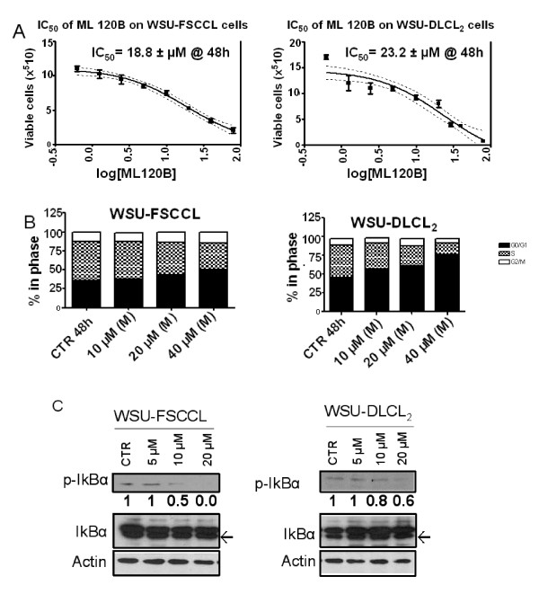
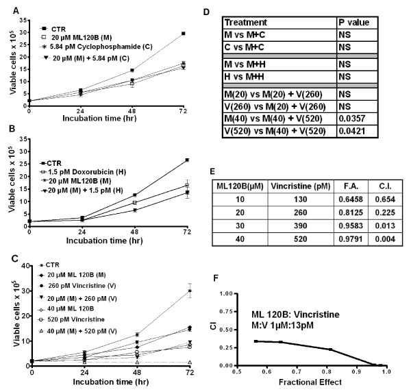
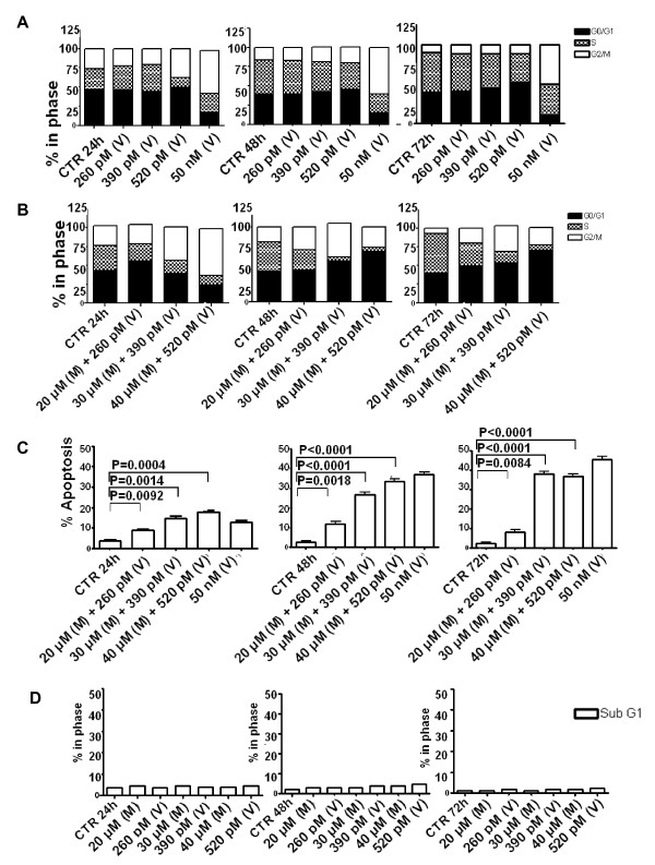
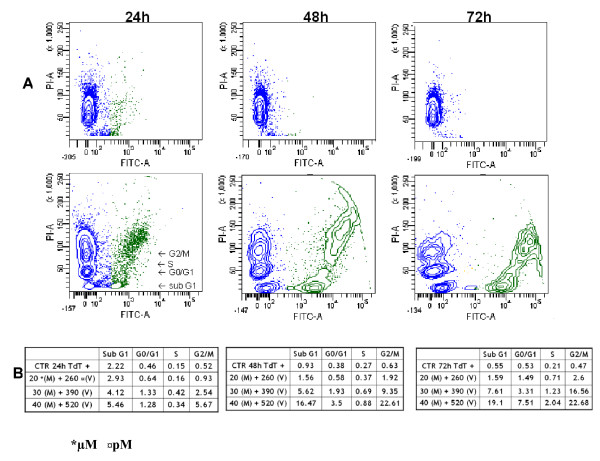
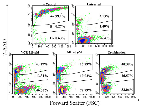
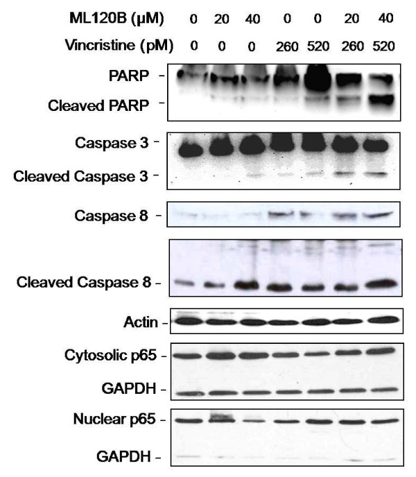
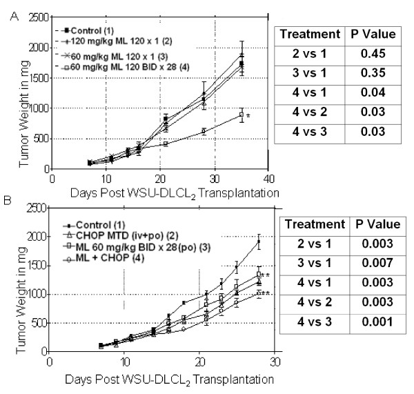
Results: ML120B down-regulated p-IκBα protein expression in a concentration dependent manner, caused growth inhibition, increased G0/G1 cells, but did not induce apoptosis. There was no significant enhancement of cell kill in the M/C or M/H combination. However, there was strong synergy in the M/V combination where the vincristine concentration can be lowered by a hundred fold in the combination for comparable G2/M arrest and apoptosis. ML120B prevented vincristine-induced nuclear translocation of p65 subunit of NF-κB. In vivo, ML120B was effective by itself and enhanced CHOP anti-tumor activity significantly (P = 0.001) in the WSU-DLCL2-SCID model but did not prevent CNS lymphoma in the WSU-FSCCL-SCID model.
Conclusions: For the first time, this study demonstrates that perturbation of IKK-2 by ML120B leads to synergistic enhancement of vincristine cytotoxicity in lymphoma. These results suggest that disruption of the NF-κB pathway is a useful adjunct to cytotoxic chemotherapy in lymphoma.
Figures
Similar articles
-
Genistein sensitizes diffuse large cell lymphoma to CHOP (cyclophosphamide, doxorubicin, vincristine, prednisone) chemotherapy.Mol Cancer Ther. 2003 Dec;2(12):1361-8. Mol Cancer Ther. 2003. PMID: 14707277
-
The addition of bryostatin 1 to cyclophosphamide, doxorubicin, vincristine, and prednisone (CHOP) chemotherapy improves response in a CHOP-resistant human diffuse large cell lymphoma xenograft model.Clin Cancer Res. 2000 Dec;6(12):4950-6. Clin Cancer Res. 2000. PMID: 11156256
-
Superior antitumor activity of SAR3419 to rituximab in xenograft models for non-Hodgkin's lymphoma.Clin Cancer Res. 2009 Jun 15;15(12):4038-45. doi: 10.1158/1078-0432.CCR-08-2808. Epub 2009 Jun 9. Clin Cancer Res. 2009. PMID: 19509168
-
Cardiovascular adverse events in patients with non-Hodgkin lymphoma treated with first-line cyclophosphamide, doxorubicin, vincristine, and prednisone (CHOP) or CHOP with rituximab (R-CHOP): a systematic review and meta-analysis.Lancet Haematol. 2020 Apr;7(4):e295-e308. doi: 10.1016/S2352-3026(20)30031-4. Epub 2020 Mar 2. Lancet Haematol. 2020. PMID: 32135128
-
The therapeutic use of rituximab in non-Hodgkin's lymphoma.Eur J Haematol Suppl. 2007 Jan;(67):5-14. doi: 10.1111/j.1600-0609.2006.00789.x. Eur J Haematol Suppl. 2007. PMID: 17206982 Review.
Cited by
-
Optimized Doxorubicin Chemotherapy for Diffuse Large B-cell Lymphoma Exploits Nanocarrier Delivery to Transferrin Receptors.Cancer Res. 2021 Feb 1;81(3):763-775. doi: 10.1158/0008-5472.CAN-20-2674. Epub 2020 Nov 11. Cancer Res. 2021. PMID: 33177062 Free PMC article.
-
PNT2258, a novel deoxyribonucleic acid inhibitor, induces cell cycle arrest and apoptosis via a distinct mechanism of action: a new class of drug for non-Hodgkin's lymphoma.Oncotarget. 2016 Jul 5;7(27):42374-42384. doi: 10.18632/oncotarget.9872. Oncotarget. 2016. PMID: 27283896 Free PMC article.
-
Targeting IKKβ in Cancer: Challenges and Opportunities for the Therapeutic Utilisation of IKKβ Inhibitors.Cells. 2018 Aug 23;7(9):115. doi: 10.3390/cells7090115. Cells. 2018. PMID: 30142927 Free PMC article. Review.
-
Toll-like receptors are potential therapeutic targets in rheumatoid arthritis.World J Biol Chem. 2011 Jul 26;2(7):167-72. doi: 10.4331/wjbc.v2.i7.167. World J Biol Chem. 2011. PMID: 21912729 Free PMC article.
-
Close Encounters of the First Kind: Innate Sensors and Multiple Sclerosis.Mol Neurobiol. 2017 Jan;54(1):101-114. doi: 10.1007/s12035-015-9665-5. Epub 2016 Jan 5. Mol Neurobiol. 2017. PMID: 26732593 Review.
References
-
- A clinical evaluation of the International Lymphoma Study Group classification of non Hodgkin's lymphoma. The Non-Hodgkin's Lymphoma Classification Project. Blood. 1997;89:3909–3918. - PubMed
-
- Tsartsidze E, Betaneli M, Sharikadze N, Shavidze N, Seskuria N. Treatment of aggressive non-Hodgkin's lymphomas. Georgian Med News. 2007;145:30–33. - PubMed
Publication types
MeSH terms
Substances
Grants and funding
LinkOut - more resources
Full Text Sources
Medical
Research Materials
