

Morphologic transformation of human breast epithelial cells MCF-10A: dependence on an oxidative microenvironment and estrogen/epidermal growth factor receptors
- PMID: 20809984
- PMCID: PMC2944135
- DOI: 10.1186/1475-2867-10-30
Morphologic transformation of human breast epithelial cells MCF-10A: dependence on an oxidative microenvironment and estrogen/epidermal growth factor receptors
Abstract
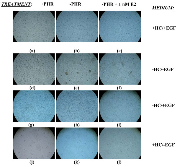
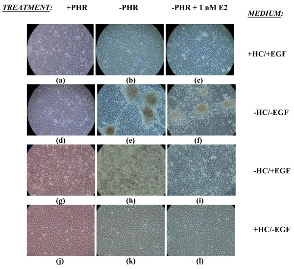
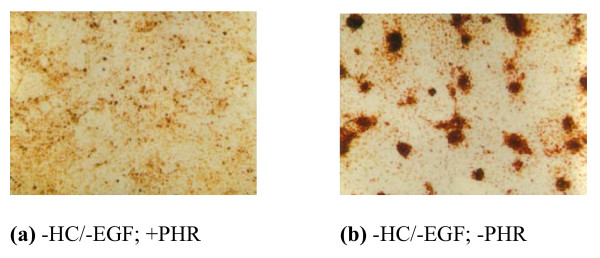
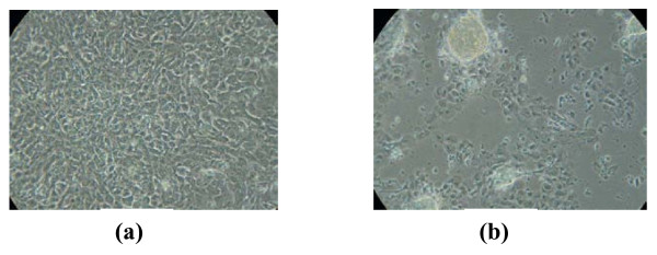
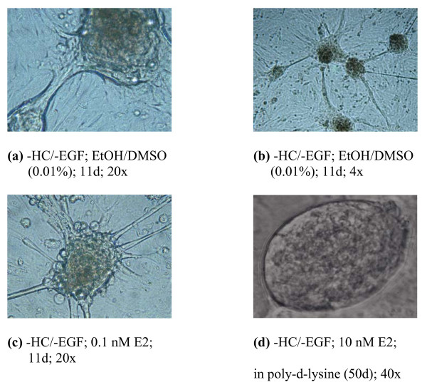
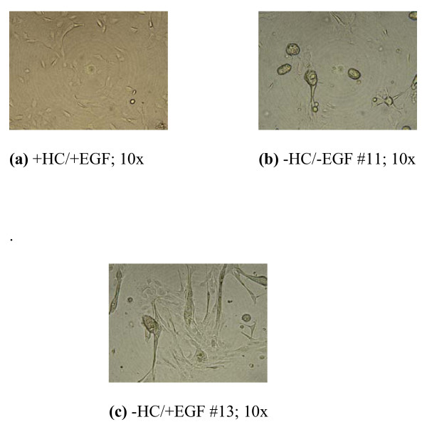
Background: MCF-10A, immortalized but non-transformed human breast epithelial cells, are widely used in research examining carcinogenesis. The studies presented here were initiated with the observation that MCF-10A cells left in continuous culture for prolonged periods without re-feeding were prone to the development of transformed foci. We hypothesized that the depletion of labile culture components led to the onset of processes culminating in the observed cell transformation. The purpose of this study was to define the factors which promoted transformation of this cell line.
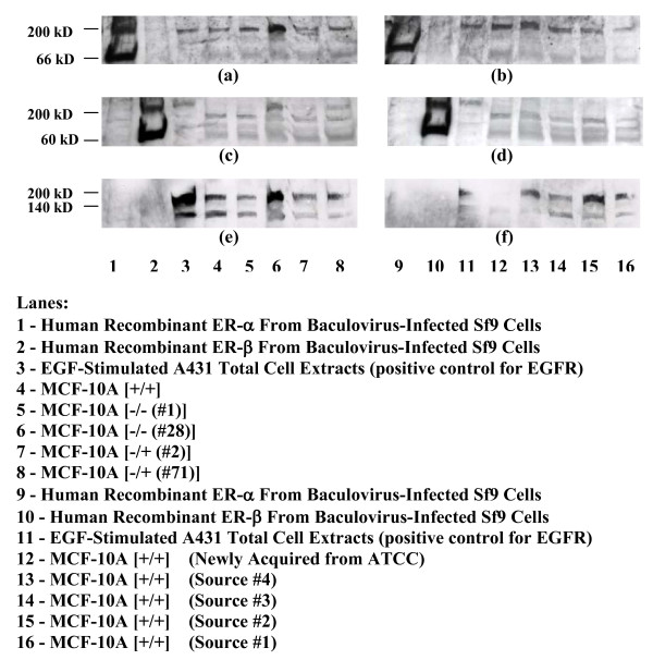
Results: Changes in levels of phenol red (PHR), hydrocortisone (HC), and epidermal growth factor (EGF) with or without estrogen treatment indicated that both oxidative stress- and estrogen receptor alpha (ERα)-mediated pathways contribute to cell transformation. Gene array and Western blotting analyses of cells maintained in our laboratory and of those from other sources documented detectable ERα and ERbeta (ERβ) in this ERα-negative cataloged cell line. Results also indicate the possibility of a direct association of EGF receptor (EGFR) and ERα in these cells as well as the formation and high induction of a novel ternary complex that includes ERβ (ERα/ERβ/EGFR) in cells grown under conditions facilitating transformation.
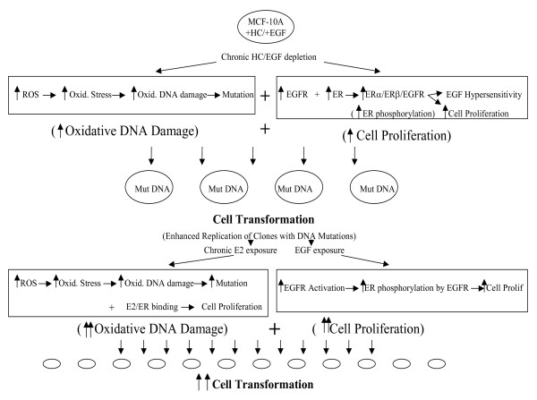
Conclusions: Our studies resulted in the development of a growth protocol where the effects of chronic, physiologically relevant alterations in the microenvironment on cellular transformation were examined. From our results, we were able to propose a model of transformation within the MCF-10A cell line in which oxidative stress, ER and EGFR play essential roles. Overall, our work indicates that the immediate microenvironment of cells exerts powerful growth cues which ultimately determine their transformation potential.
Figures
Similar articles
-
Aromatase overexpression induces malignant changes in estrogen receptor α negative MCF-10A cells.Oncogene. 2013 Oct 31;32(44):5233-40. doi: 10.1038/onc.2012.558. Epub 2012 Nov 26. Oncogene. 2013. PMID: 23178495
-
Estrogen receptor beta exerts growth-inhibitory effects on human mammary epithelial cells.Breast Cancer Res Treat. 2010 Apr;120(3):557-65. doi: 10.1007/s10549-009-0413-2. Epub 2009 May 12. Breast Cancer Res Treat. 2010. PMID: 19434490
-
Transforming growth factor-alpha expression is enhanced in human mammary epithelial cells transformed by an activated c-Ha-ras protooncogene but not by the c-neu protooncogene, and overexpression of the transforming growth factor-alpha complementary DNA leads to transformation.Cell Growth Differ. 1990 Sep;1(9):407-20. Cell Growth Differ. 1990. PMID: 1981145
-
Estrogen and its metabolites are carcinogenic agents in human breast epithelial cells.J Steroid Biochem Mol Biol. 2003 Oct;87(1):1-25. doi: 10.1016/s0960-0760(03)00390-x. J Steroid Biochem Mol Biol. 2003. PMID: 14630087 Review.
-
Estrogen receptor alpha negative breast cancer patients: estrogen receptor beta as a therapeutic target.J Steroid Biochem Mol Biol. 2008 Mar;109(1-2):1-10. doi: 10.1016/j.jsbmb.2007.12.010. Epub 2007 Dec 8. J Steroid Biochem Mol Biol. 2008. PMID: 18243688 Review.
Cited by
-
Development and characterisation of a 3D multi-cellular in vitro model of normal human breast: a tool for cancer initiation studies.Oncotarget. 2015 May 30;6(15):13731-41. doi: 10.18632/oncotarget.3803. Oncotarget. 2015. PMID: 25915532 Free PMC article.
-
Runx1 stabilizes the mammary epithelial cell phenotype and prevents epithelial to mesenchymal transition.Oncotarget. 2017 Mar 14;8(11):17610-17627. doi: 10.18632/oncotarget.15381. Oncotarget. 2017. PMID: 28407681 Free PMC article.
-
Role of thioredoxin reductase 1 in dysplastic transformation of human breast epithelial cells triggered by chronic oxidative stress.Sci Rep. 2016 Nov 15;6:36860. doi: 10.1038/srep36860. Sci Rep. 2016. PMID: 27845427 Free PMC article.
-
Reactive oxygen species via redox signaling to PI3K/AKT pathway contribute to the malignant growth of 4-hydroxy estradiol-transformed mammary epithelial cells.PLoS One. 2013;8(2):e54206. doi: 10.1371/journal.pone.0054206. Epub 2013 Feb 21. PLoS One. 2013. PMID: 23437041 Free PMC article.
-
IL6-mediated suppression of miR-200c directs constitutive activation of inflammatory signaling circuit driving transformation and tumorigenesis.Mol Cell. 2012 Mar 30;45(6):777-89. doi: 10.1016/j.molcel.2012.01.015. Epub 2012 Feb 23. Mol Cell. 2012. PMID: 22364742 Free PMC article.
References
-
- Gullick JW, Bianco C, Normanno N, Martinez-Lacacia I, De Santis M, Ebert AD, Salomon DS. Growth factors and their receptors: a novel approach to the endocrinology of human breast cancer. Women and Cancer. 1998;1:29–57.
-
- Ohshima H, Bartsche H. Chronic infections and inflammatory processes as cancer risk factors: possible role of nitric oxide in carcinogenesis. Mutation Res. 1994;305:253–256. - PubMed
-
- Yager JD. Endogenous estrogens as carcinogens through metabolic activation. J Natl Cancer Inst. 2000;27:67–73. - PubMed
-
- Hayashi N, Hasegawa K, Komine A, Tanaka Y, McLachlan JA, Barrett JC, Tsutsui T. Estrogen-induced cell transformation and DNA adduct formation in cultured Syrian hamster embryo cells. Mol Carcinogenesis. 1996;16:149–156. doi: 10.1002/(SICI)1098-2744(199607)16:3<149::AID-MC5>3.0.CO;2-C. - DOI - PubMed
LinkOut - more resources
Full Text Sources
Research Materials
Miscellaneous
