

Toward implementing an MRI-based PET attenuation-correction method for neurologic studies on the MR-PET brain prototype
- PMID: 20810759
- PMCID: PMC3801218
- DOI: 10.2967/jnumed.109.069112
Toward implementing an MRI-based PET attenuation-correction method for neurologic studies on the MR-PET brain prototype
Abstract
Several factors have to be considered for implementing an accurate attenuation-correction (AC) method in a combined MR-PET scanner. In this work, some of these challenges were investigated, and an AC method based entirely on the MRI data obtained with a single dedicated sequence was developed and used for neurologic studies performed with the MR-PET human brain scanner prototype.
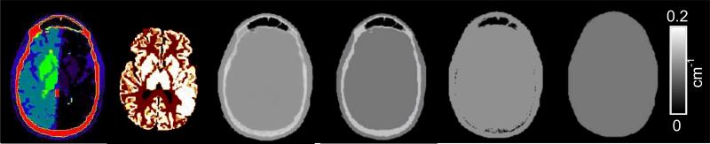
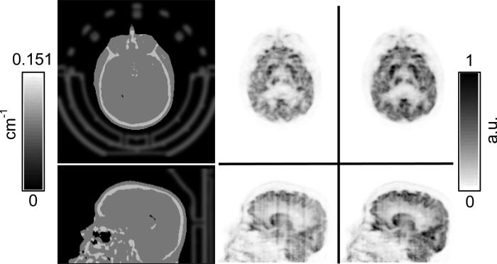
Methods: The focus was on the problem of bone-air segmentation, selection of the linear attenuation coefficient for bone, and positioning of the radiofrequency coil. The impact of these factors on PET data quantification was studied in simulations and experimental measurements performed on the combined MR-PET scanner. A novel dual-echo ultrashort echo time (DUTE) MRI sequence was proposed for head imaging. Simultaneous MR-PET data were acquired, and the PET images reconstructed using the proposed DUTE MRI-based AC method were compared with the PET images that had been reconstructed using a CT-based AC method.
Results: Our data suggest that incorrectly accounting for the bone tissue attenuation can lead to large underestimations (>20%) of the radiotracer concentration in the cortex. Assigning a linear attenuation coefficient of 0.143 or 0.151 cm(-1) to bone tissue appears to give the best trade-off between bias and variability in the resulting images. Not identifying the internal air cavities introduces large overestimations (>20%) in adjacent structures. On the basis of these results, the segmented CT AC method was established as the silver standard for the segmented MRI-based AC method. For an integrated MR-PET scanner, in particular, ignoring the radiofrequency coil attenuation can cause large underestimations (i.e., <or=50%) in the reconstructed images. Furthermore, the coil location in the PET field of view has to be accurately known. High-quality bone-air segmentation can be performed using the DUTE data. The PET images obtained using the DUTE MRI- and CT-based AC methods compare favorably in most of the brain structures.
Conclusion: A DUTE MRI-based AC method considering all these factors was implemented. Preliminary results suggest that this method could potentially be as accurate as the segmented CT method and could be used for quantitative neurologic MR-PET studies.
Figures


References
-
- Judenhofer MS, Wehrl HF, Newport DF, et al. Simultaneous PET-MRI: a new approach for functional and morphological imaging. Nat Med. 2008 Apr;14(4):459–465. - PubMed
-
- Lucas AJ, Hawkes RC, Ansorge RE, et al. Development of a combined microPET((R))-MR system. Technol Cancer Res Treat. 2006 Aug;5(4):337–341. - PubMed
-
- Woody C, Schlyer D, Vaska P, et al. Preliminary studies of a simultaneous PET/MRI scanner based on the RatCAP small animal tomograph. Nucl Instrum Methods Phys Res Sect A Accel Spectrom Dect Assoc Equip. 2007 Feb;571(1-2):102–105.
Publication types
MeSH terms
Grants and funding
LinkOut - more resources
Full Text Sources
Other Literature Sources
Medical