

The effect of skeletal maturity on functional healing of the anterior cruciate ligament
- PMID: 20810854
- PMCID: PMC2924734
- DOI: 10.2106/JBJS.I.01368
The effect of skeletal maturity on functional healing of the anterior cruciate ligament
Abstract
Background: The effects of skeletal maturity on functional ligament healing are unknown. Prior studies have suggested that ligament injuries in skeletally mature animals heal with improved mechanical properties. In this study, we hypothesized that skeletally immature animals have improved functional healing compared with skeletally mature animals.
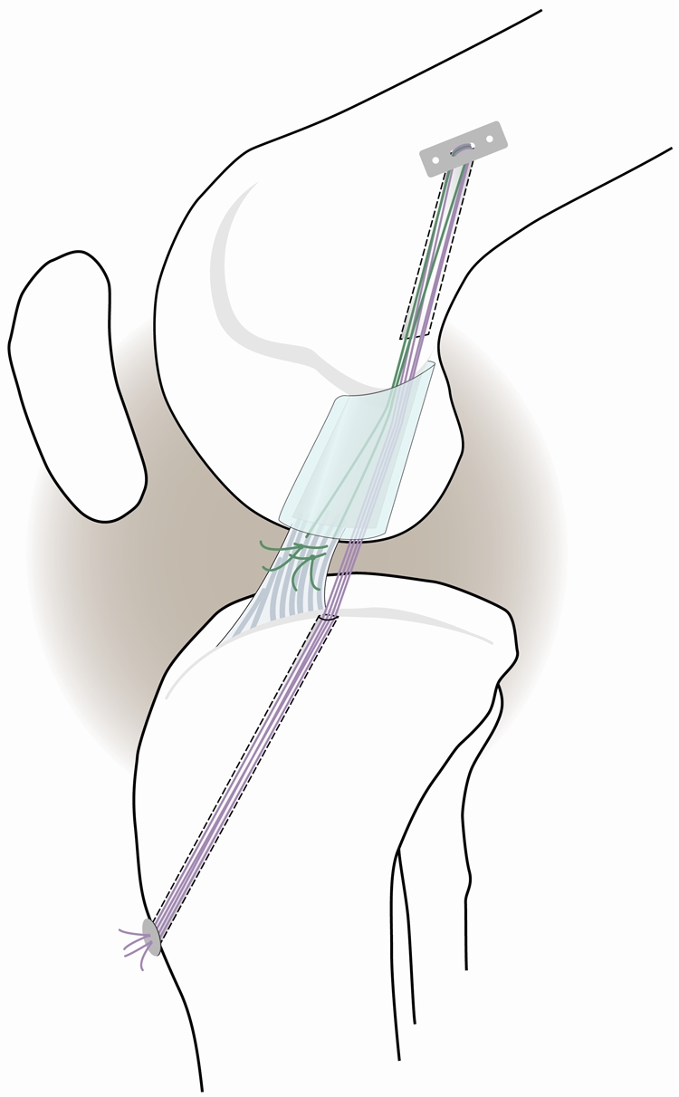
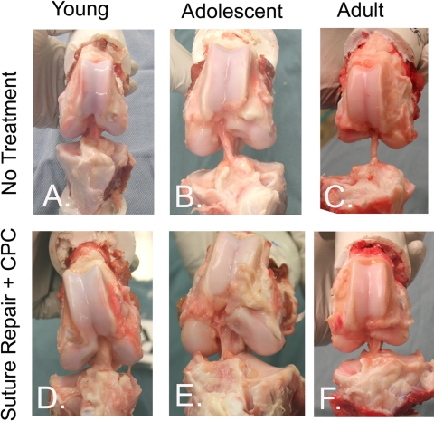
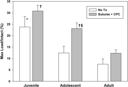
Methods: Twenty-one Yucatan minipigs (eight juvenile, eight adolescent, and five adult animals) underwent bilateral anterior cruciate ligament transection. On one side, the ligament injury was left untreated to determine the intrinsic healing response as a function of age. On the contralateral side, an enhanced suture repair incorporating a collagen-platelet composite was performed. Biomechanical properties of the repairs were measured after fifteen weeks of healing, and histologic analysis was performed.
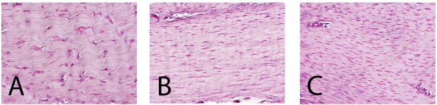
Results: Anterior cruciate ligaments from skeletally immature animals had significantly improved structural properties over those of adult animals at three months after transection in both the untreated and repair groups. Use of the enhanced suture technique provided the most improvement in the adolescent group, in which an increase of 85% in maximum load was noted with repair. The repair tissue in the adult tissue had the highest degree of hypercellularity at the fifteen-week time point.
Conclusions: Functional ligament healing depends on the level of skeletal maturity of the animal, with immature animals having a more productive healing response than mature animals.
Figures
Comment in
-
Commentary on an article by Martha M. Murray, MD, et al.: "The effect of skeletal maturity on functional healing of the anterior cruciate ligament".J Bone Joint Surg Am. 2010 Sep 1;92(11):e15. doi: 10.2106/JBJS.J.00940. J Bone Joint Surg Am. 2010. PMID: 20810851 No abstract available.
References
-
- Murray MM, Martin SD, Martin TL, Spector M. Histological changes in the human anterior cruciate ligament after rupture. J Bone Joint Surg Am. 2000;82:1387-97 - PubMed
-
- Murray MM, Spindler KP, Ballard P, Welch TP, Zurakowski D, Nanney LB. Enhanced histologic repair in a central wound in the anterior cruciate ligament with a collagen-platelet-rich plasma scaffold. J Orthop Res. 2007;25:1007-17 - PubMed
-
- Murray MM, Spindler KP, Devin C, Snyder BS, Muller J, Takahashi M, Ballard P, Nanney LB, Zurakowski D. Use of a collagen-platelet rich plasma scaffold to stimulate healing of a central defect in the canine ACL. J Orthop Res. 2006;24:820-30 - PubMed
Publication types
MeSH terms
Grants and funding
LinkOut - more resources
Full Text Sources
