. 2010 Dec;299(6):C1468-84.
doi: 10.1152/ajpcell.00246.2010. Epub 2010 Sep 1.

PECAM-1 regulates proangiogenic properties of endothelial cells through modulation of cell-cell and cell-matrix interactions

Affiliations

PECAM-1 regulates proangiogenic properties of endothelial cells through modulation of cell-cell and cell-matrix interactions

SunYoung Park et al. Am J Physiol Cell Physiol. 2010 Dec.

Abstract

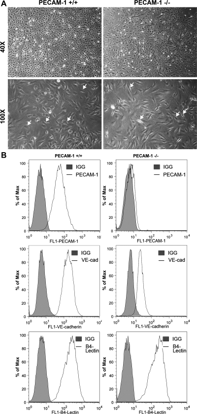
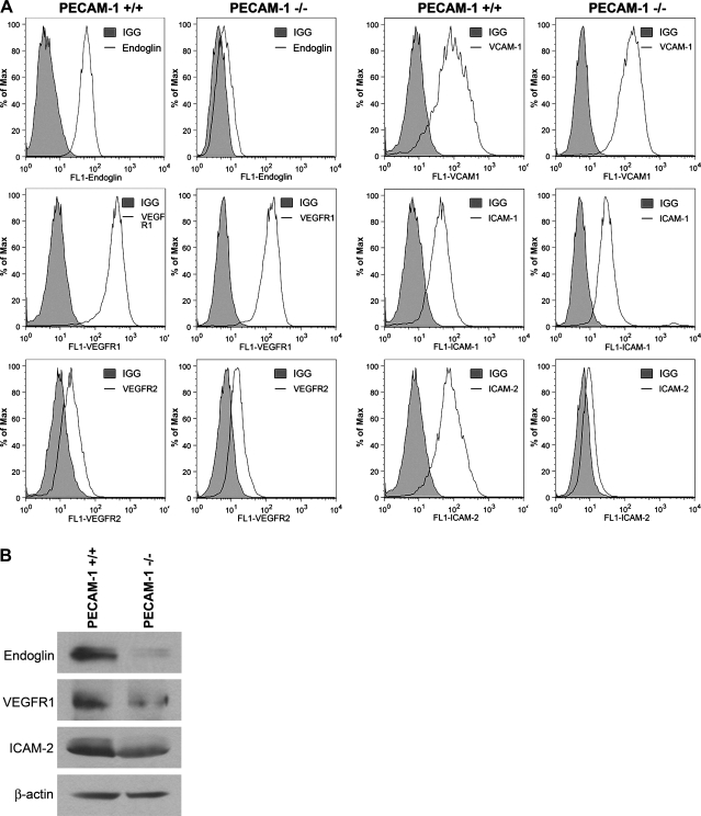
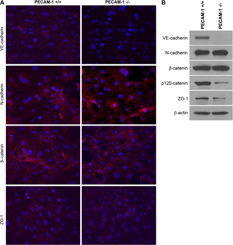
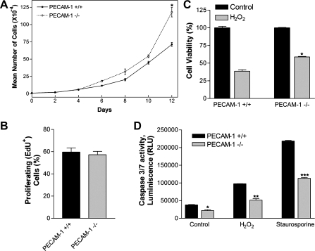
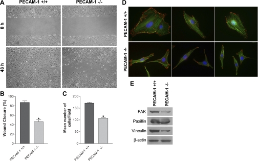
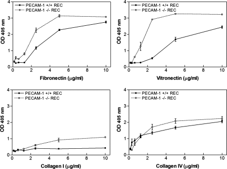
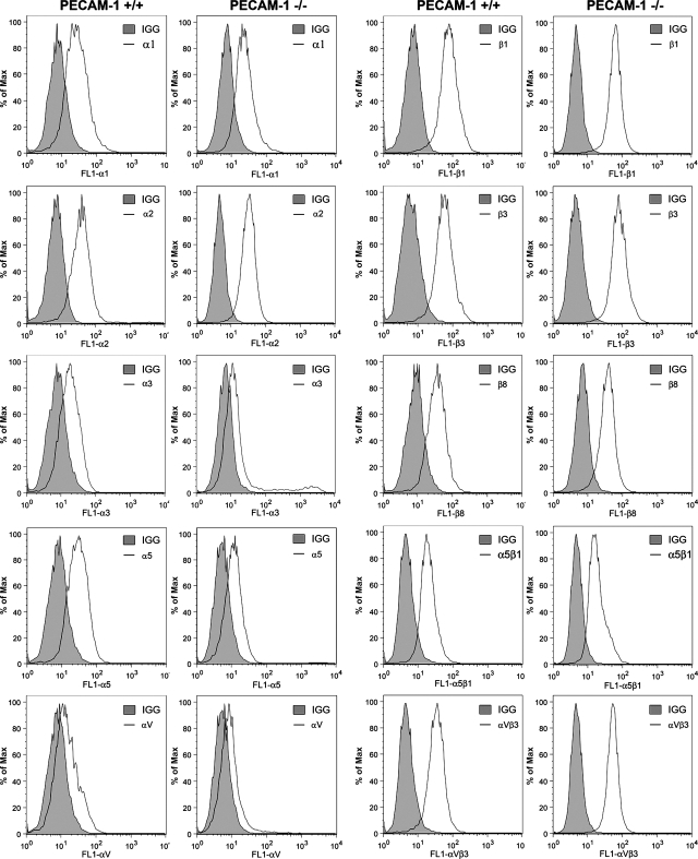
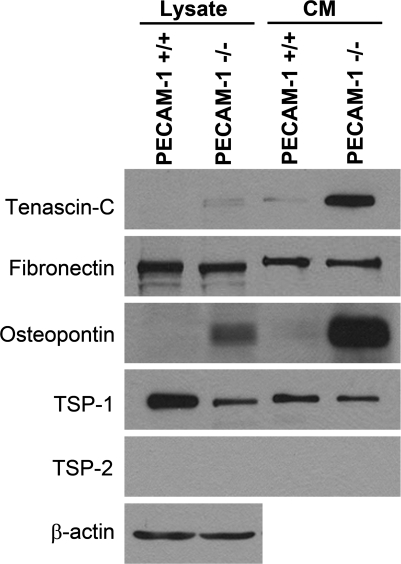
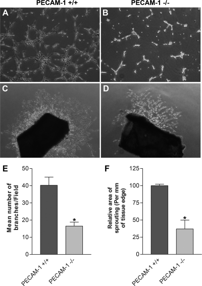
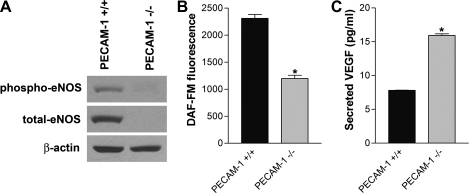
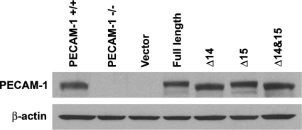
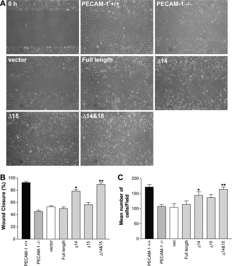
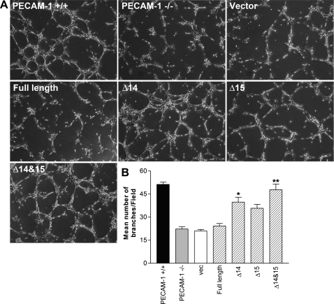
Platelet endothelial cell adhesion molecule-1 (PECAM-1/CD31) is a member of the immunoglobulin superfamily of cell adhesion molecules with important roles in angiogenesis and inflammation. However, the molecular and cellular mechanisms, and the role that specific PECAM-1 isoforms play in these processes, remain elusive. We recently showed attenuation of retinal vascular development and neovascularization in PECAM-1-deficient (PECAM-1-/-) mice. To gain further insight into the role of PECAM-1 in these processes, we isolated primary retinal endothelial cells (EC) from wild-type (PECAM-1+/+) and PECAM-1-/- mice. Lack of PECAM-1 had a significant impact on endothelial cell-cell and cell-matrix interactions, resulting in attenuation of cell migration and capillary morphogenesis. Mechanistically these changes were associated with a significant decrease in expression of endothelial nitric oxide synthase (eNOS) and nitric oxide (NO) bioavailability in PECAM-1-/- retinal EC. PECAM-1-/- retinal EC also exhibited a lower rate of apoptosis under basal and challenged conditions, consistent with their increased growth rate. Furthermore, reexpression of PECAM-1 was sufficient to restore migration and capillary morphogenesis of null cells in an isoform-specific manner. Thus PECAM-1 expression modulates proangiogenic properties of EC, and these activities are significantly influenced by alternative splicing of its cytoplasmic domain.


Figures

Fig. 1.
Fig. 1.
Isolation and characterization of mouse retinal endothelial cells (EC). Platelet endothelial cell adhesion molecule (PECAM)-1+/+ and PECAM-1−/− retinal EC were prepared as described in materials and methods and cultured on gelatin-coated plates in 60-mm dishes. A: cells were photographed in digital format at ×40 and ×100 magnification. Note the elongated, spindly morphology of PECAM-1−/− cells compared with PECAM-1+/+ cells (arrows). B: expression of vascular EC markers in EC. Retinal EC were examined for expression of PECAM-1, VE-cadherin (VE-cad), and B4 lectin by FACS analysis. Shaded areas show control IgG staining. These experiments were repeated with 3 isolations of EC, with similar results.
Fig. 2.
Fig. 2.
Expression of other EC markers in PECAM-1+/+ and PECAM-1−/− retinal EC. A: expression of endoglin, VEGF receptor (VEGFR)1, VEGFR2, VCAM-1, ICAM-1, and ICAM-2 was determined by FACS analysis of PECAM-1+/+ and PECAM-1−/− retinal EC using specific antibodies. Shaded areas show control IgG staining. Note decreased expression of endoglin, VEGFR1, and ICAM-2 in PECAM-1−/− EC. B: changes in the levels of these proteins were further confirmed by Western blot analysis. These experiments were repeated with 3 isolations of EC, with similar results.
Fig. 3.
Fig. 3.
Cellular localization and expression level of VE-cadherin, N-cadherin, β-catenin, and ZO-1. A: PECAM-1+/+ and PECAM-1−/− retinal EC were grown on fibronectin-coated chamber slides to confluence and stained as described in materials and methods with specific antibodies. No staining was observed when primary antibody was left out. Note reduced expression of VE-cadherin and ZO-1 and their absence from the sites of cell-cell contact. Although the levels of N-cadherin were comparable, the junctional localization of N-cadherin was more prominent in PECAM-1−/− retinal EC compared with PECAM-1+/+ EC. B: Western blot analysis of cell lysates for junctional proteins. Cell lysates prepared from PECAM-1+/+ and PECAM-1−/− retinal EC were analyzed for expression of different junction proteins including VE-cadherin, N-cadherin, β-catenin, p120-catenin, ZO-1, and β-actin by Western blotting. Note significant decrease in protein levels of VE-cadherin and ZO-1 in PECAM-1−/− retinal EC. The levels of β-catenin and actin were similar. These experiments were repeated twice with 2 isolations of these cells, with similar results.
Fig. 4.
Fig. 4.
Altered proliferation and apoptosis of PECAM-1−/− retinal EC. A and B: the rate of retinal EC proliferation was determined by counting the number of cells in triplicate after different days in culture as described in materials and methods (A) and by analyzing the rate of DNA synthesis by FACScan flow cytometer analysis (B; P > 0.05; n = 4). EdU, 5-ethynyl-2′-deoxyuridine. C: hydrogen peroxide (H2O2) toxicity of retinal EC was measured with the 3-(4,5-dimethylthiazol-2-yl)-5-(3-carboxymethoxyphenyl)-2-(4-sulfophenyl)-2H-tetrazolium (MTS) assay method. Retinal EC were incubated with 1.75 mM H2O2 in EC growth medium for 2 days, and the level of toxicity was determined by measuring colorimetric change of MTS in cells on 96-well plates. PECAM-1−/− retinal EC were significantly less sensitive to cytotoxic effect of H2O2 (*P < 0.05; n = 5). D: rate of apoptosis was determined by measuring caspase activity with luminescent signal from caspase-3/7 DEVD-aminoluciferin substrate, as recommended by the supplier. As an apoptotic stimulus, H2O2 and staurosporine in EC growth media were added for 8 h. Note the significant decrease in the rate of apoptosis in PECAM-1−/− retinal EC compared with PECAM-1+/+ retinal EC (*,**,***P < 0.05; n = 5). RLU, relative light units.
Fig. 5.
Fig. 5.
PECAM-1−/− retinal EC are less migratory. A: cell migration was determined by scratch wound assay of the retinal EC monolayer on gelatin-coated plates. Wound closure was monitored by photography within 48 h. B: quantitative assessment of the data (*P < 0.05, n = 3). C: rate of cell migration was also determined with a Transwell migration assay (*P < 0.05, n = 3). D: the migratory phenotype of retinal EC was further confirmed by staining the cells with phalloidin (green, actin) and vinculin (red, focal adhesions). Note increased actin stress fibers and focal adhesions in PECAM-1+/+ cells. Fewer focal adhesions were detected in PECAM-1−/− cells, which mainly localized to the basal surface of the cells. E: decreased expression of focal adhesion proteins including focal adhesion kinase (FAK), paxillin, and vinculin was detected in PECAM-1−/− EC. β-Actin was used as loading control. These experiments were repeated with 3 isolations of cells, with similar results. Note the flatter morphology and peripheral actin localization in PECAM-1−/− EC.
Fig. 6.
Fig. 6.
Enhanced adhesion of PECAM-1−/− retinal EC (REC) to various extracellular matrix (ECM) proteins. Adhesion of retinal EC to fibronectin, vitronectin, collagen I, and collagen IV was determined as described in materials and methods. Note strong adhesion of PECAM-1−/− retinal EC to fibronectin and vitronectin. These experiments were performed at least twice with 2 different isolations of retinal EC. OD, optical density.
Fig. 7.
Fig. 7.
Expression of integrins in retinal EC. α1-, α2, α3-, α5-, αv-, β1-, β3-, β8-, α5β1-, and αvβ3-integrin expression on retinal EC was determined by FACS analysis as described in materials and methods. These experiments were repeated with 2 isolations of cells, with similar results. Note similar expression of integrins in both cell types.
Fig. 8.
Fig. 8.
Altered expression of ECM proteins in PECAM-1−/− retinal EC. Retinal EC from PECAM-1+/+ and PECAM-1−/− mice were incubated for 2 days in serum-free medium. The collected conditioned medium (CM) and cell lysates were analyzed by Western blotting for tenascin-C, fibronectin, osteopontin, thrombospondin (TSP)1, and TSP2 with specific antibodies. These experiments were repeated with 2 different isolations, with similar results. Note significant upregulation of tenascin-C and osteopontin in PECAM-1−/− retinal EC.
Fig. 9.
Fig. 9.
Attenuation of capillary morphogenesis of retinal EC and sprouting of aortas from PECAM-1−/− mice. A and B: PECAM-1+/+ (A) and PECAM-1−/− (B) retinal EC were plated on Matrigel, and capillary morphogenesis was monitored for 3 days. The photographs were taken in digital format after 18 h when optimal capillary morphogenesis was observed. C and D: aortas prepared from PECAM-1+/+ (C) and PECAM-1−/− (D) mice were cultured on Matrigel, and sprouting was monitored for 6 days and photographed in digital format. Quantitative assessments of the data are shown in E and F, respectively. Data are the mean number of branch points from 5 high-power fields (×100). Note a significant decrease in capillary morphogenesis of PECAM-1−/− EC compared with PECAM-1+/+ cells (*P < 0.05; n = 5). Also note a significant decrease in sprouting of aortas from PECAM-1−/− mice compared with PECAM-1+/+ mice (*P < 0.05; n = 6). These experiments were repeated with 3 different isolations of retinal EC and aortas from 5 different mice, with similar results.
Fig. 10.
Fig. 10.
Altered expression and phosphorylation of endothelial nitric oxide synthase (eNOS) and VEGF expression in PECAM-1−/− retinal EC. A: phospho-eNOS and total eNOS in cell lysates were analyzed by Western blotting. β-Actin was used for loading control. B: intracellular nitric oxide (NO) level in retinal EC was measured with 4-amino-5-methylamino-2,7-difluorofluorescein (DAF-FM) as described in materials and methods. Note a significant decrease in intracellular NO level in PECAM-1−/− retinal EC compared with PECAM-1+/+ retinal EC (*P < 0.05; n = 3). C: secreted level of VEGF in retinal EC was determined with an immunoassay as described in materials and methods. Note a significant increase in the level of VEGF secreted by PECAM-1−/− retinal EC compared with PECAM-1+/+ EC (*P < 0.05; n = 3). These experiments were repeated with 3 isolations of cells, with similar results.
Fig. 11.
Fig. 11.
Expression of various PECAM-1 isoforms in PECAM-1−/− retinal EC. Adenoviruses encoding empty vector or a specific PECAM-1 isoform [full length or isoform lacking exon 14 (Δ14), 15 (Δ15), or 14 and 15 (Δ14&15)] were used to infect PECAM-1−/− retinal EC as described in materials and methods. The level of PECAM-1 was determined by Western blotting of cell lysates. Note similar expression level of PECAM-1 in PECAM-1−/− retinal EC compared with PECAM-1+/+ retinal EC.
Fig. 12.
Fig. 12.
Reexpression of PECAM-1 restored migration of PECAM-1−/− retinal EC in an isoform-specific manner. A: cell migration was determined by scratch wound assay of monolayer of retinal EC expressing the empty vector or a specific PECAM-1 isoform. Wound closure was monitored by photography within 48 h and quantified relative to time zero. B: quantitative assessment of the data (*,**P < 0.05, n = 3). C: similar results were observed with a Transwell migration assay (*,**P < 0.05, n = 3).
Fig. 13.
Fig. 13.
Reexpression of PECAM-1 restored capillary morphogenesis of PECAM-1−/− retinal EC in an isoform-specific manner. A: capillary morphogenesis was determined by plating retinal EC expressing the empty vector or a specific PECAM-1 isoform on Matrigel and photographed after 18 h. B: quantitative assessment of data. Data are the mean number of branch points from 5 high-power fields (×100). Note the significant restoration of capillary morphogenesis in PECAM-1−/− retinal EC expressing Δ14&15 PECAM-1 isoform. These experiments were repeated with 2 different isolations of retinal EC. *,**P < 0.05; n = 3.

References

    1. Alderton WK, Cooper CE, Knowles RG. Nitric oxide synthases: structure, function and inhibition. Biochem J 357: 593– 615, 2001 - PMC - PubMed
    1. Bagi Z, Frangos JA, Yeh JC, White CR, Kaley G, Koller A. PECAM-1 mediates NO-dependent dilation of arterioles to high temporal gradients of shear stress. Arterioscler Thromb Vasc Biol 25: 1590– 1595, 2005 - PubMed
    1. Bergom C, Paddock C, Gao C, Holyst T, Newman DK, Newman PJ. An alternatively spliced isoform of PECAM-1 is expressed at high levels in human and murine tissues, and suggests a novel role for the C-terminus of PECAM-1 in cytoprotective signaling. J Cell Sci 121: 1235– 1242, 2008 - PMC - PubMed
    1. Carmeliet P, Lampugnani MG, Moons L, Breviario F, Compernolle V, Bono F, Balconi G, Spagnuolo R, Oostuyse B, Dewerchin M, Zanetti A, Angellilo A, Mattot V, Nuyens D, Lutgens E, Clotman F, de Ruiter MC, Gittenberger-de Groot A, Poelmann R, Lupu F, Herbert JM, Collen D, Dejana E. Targeted deficiency or cytosolic truncation of the VE-cadherin gene in mice impairs VEGF-mediated endothelial survival and angiogenesis. Cell 98: 147– 157, 1999 - PubMed
    1. Cheresh DA, Stupack DG. Regulation of angiogenesis: apoptotic cues from the ECM. Oncogene 27: 6285– 6298, 2008 - PubMed

Publication types

MeSH terms

Substances

LinkOut - more resources