Review
. 2010 Sep;18(9):520-7.
doi: 10.5435/00124635-201009000-00003.

Noncontact anterior cruciate ligament injuries: mechanisms and risk factors

Affiliations
Review

Noncontact anterior cruciate ligament injuries: mechanisms and risk factors

Barry P Boden et al. J Am Acad Orthop Surg. 2010 Sep.

Abstract

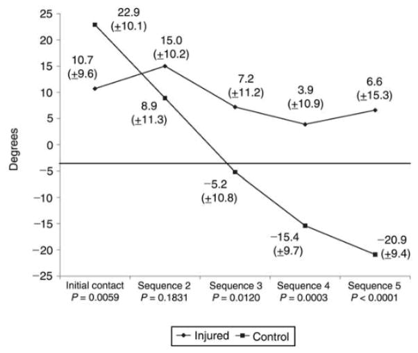
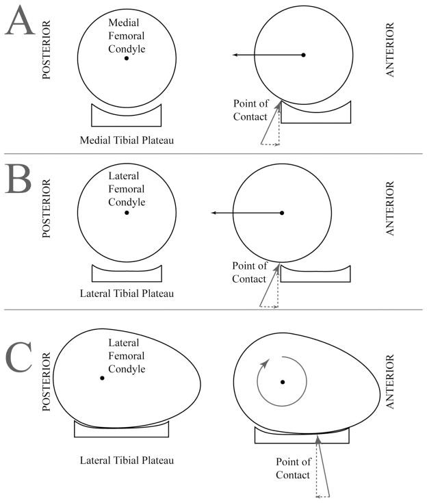
Significant advances have recently been made in understanding the mechanisms involved in noncontact anterior cruciate ligament (ACL) injury. Most ACL injuries involve minimal to no contact. Female athletes sustain a two- to eightfold greater rate of injury than do their male counterparts. Recent videotape analyses demonstrate significant differences in average leg and trunk positions during injury compared with control subjects. These findings as well as those of cadaveric and MRI studies indicate that axial compressive forces are a critical component in noncontact ACL injury. A complete understanding of the forces and risk factors associated with noncontact ACL injury should lead to the development of improved preventive strategies for this devastating injury.


Figures

Figure 1
Figure 1
Sagittal magnetic resonance image with the knee at an angle of 5°, demonstrating the angle of the patellar tendon (solid white arrow) at its attachment to the tibial tubercle. Because the angle is low (<45°), the compressive vector (PTC) is larger than the anterior shear vector (PTAS). ACL = anterior cruciate ligament.
Figure 2
Figure 2
Initial foot contact with the ground in a safe (A) and an injured (B) athlete demonstrating safe (A) and dangerous (B) landing posture. (Reproduced with permission from Boden BP, Torg JS, Knowles SB, Hewett TE: Video analysis of anterior cruciate ligament injury: Abnormalities in hip and ankle kinematics. Am J Sports Med 2009;37:252–259.)
Figure 3
Figure 3
Sagittal ankle angles in injured and uninjured athletes for the first five frames of videotape analysis, beginning with initial ground contact (30 Hz). (Reproduced with permission from Boden BP, Torg JS, Knowles SB, Hewett TE: Video analysis of anterior cruciate ligament injury: Abnormalities in hip and ankle kinematics. Am J Sports Med 2009;37:252–259.)
Figure 4
Figure 4
Illustration of the tibial plateau as the leg transitions from a safe to a provocative position. The tibial plateau has a more vertical orientation in the provocative position. (Adapted with permission from Boden BP, Breit I, Sheehan FT: Tibiofemoral alignment: Contributing factors to noncontact anterior cruciate ligament injury. J Bone Joint Surg Am 2009;91:2381–2389.)
Figure 5
Figure 5
A, Schematic representation of the medial compartment, demonstrating the concave shape of the medial tibial plateau. Left, This shape enables the femur to fit with the tibia as a ball fits into a cup. Right, The femur has the potential to slide relative to the tibia, but it runs up against the cup, so it tends to roll instead. The femur also needs to go uphill, which costs energy. B, The cup is flatter on the lateral tibial plateau (left), resulting in lesser forces when the femur tries to slide (right). The assumption that the lateral femoral condyle is circular is incorrect. C, In full extension, the anterior portion of the lateral femoral condyle, which is much flatter than the posterior aspect, is in contact with the tibial plateau (left). Thus, there are two “flat” surfaces riding against each other. For the femur to roll on the tibia, its posterior side must rise, which costs energy. Thus, it is more likely to slide (right).

References

    1. Boden BP, Dean GS, Feagin JA, Jr, Garrett WE., Jr Mechanisms of anterior cruciate ligament injury. Orthopedics. 2000;23:573–578. - PubMed
    1. Uhorchak JM, Scoville CR, Williams GN, Arciero RA, St PP, Taylor DC. Risk factors associated with noncontact injury of the anterior cruciate ligament: a prospective four-year evaluation of 859 West Point cadets. Am J Sports Med. 2003;31:831–842. - PubMed
    1. DeMorat G, Weinhold P, Blackburn T, Chudik S, Garrett W. Aggressive quadriceps loading can induce noncontact anterior cruciate ligament injury. Am J Sports Med. 2004;32:477–483. - PubMed
    1. Meyer EG, Baumer TG, Slade JM, Smith WE, Haut RC. Tibiofemoral contact pressures and osteochondral microtrauma during anterior cruciate ligament rupture due to excessive compressive loading and internal torque of the human knee. Am J Sports Med. 2008;36:1966–1977. - PubMed
    1. Boden BP, Torg JS, Knowles SB, Hewett TE. Video analysis of anterior cruciate ligament injury: abnormalities in hip and ankle kinematics. Am J Sports Med. 2009;37:252–259. - PubMed

Publication types