. 2010 Oct 1;185(7):4393-400.
doi: 10.4049/jimmunol.0903239. Epub 2010 Sep 1.

B cell subsets contribute to renal injury and renal protection after ischemia/reperfusion

Affiliations

B cell subsets contribute to renal injury and renal protection after ischemia/reperfusion

Brandon Renner et al. J Immunol. .

Abstract

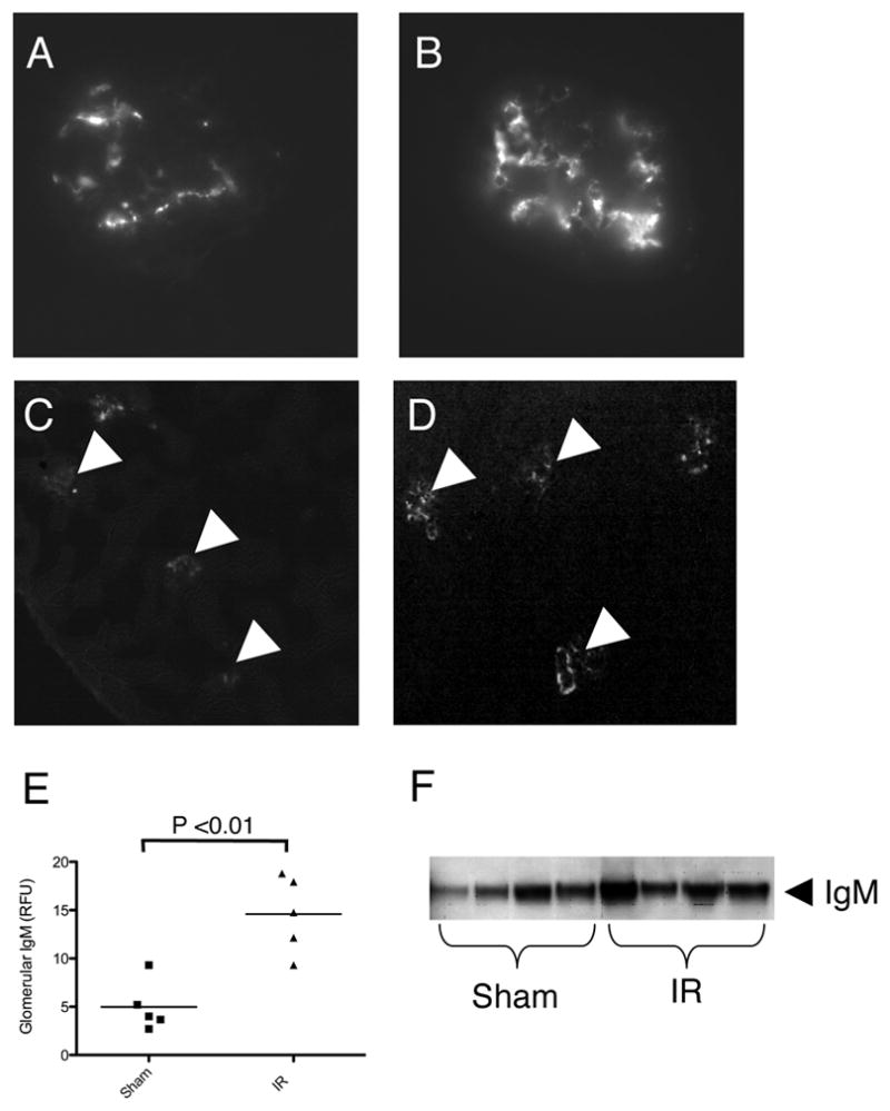
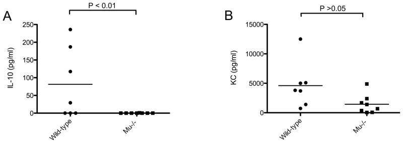
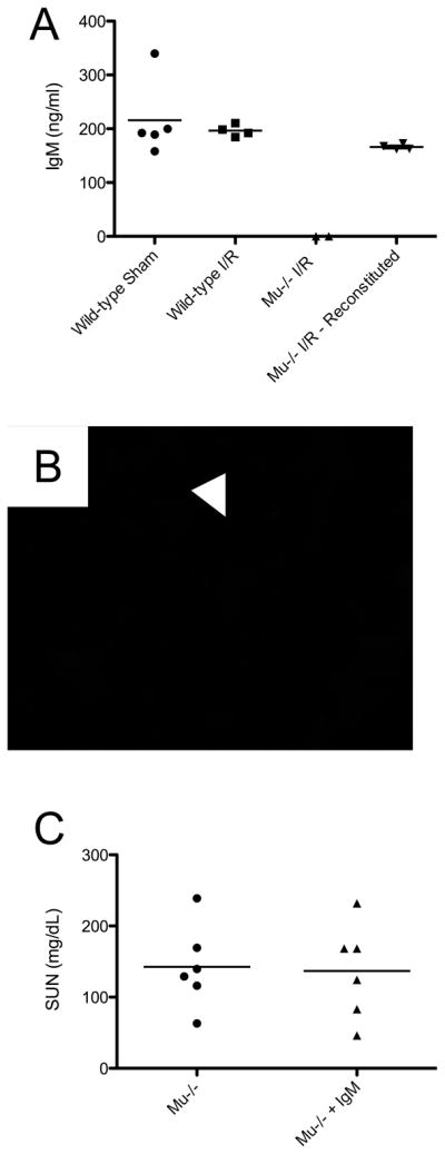
Ischemia/reperfusion (I/R) triggers a robust inflammatory response within the kidney. Numerous components of the immune system contribute to the resultant renal injury, including the complement system. We sought to identify whether natural Abs bind to the postischemic kidney and contribute to complement activation after I/R. We depleted peritoneal B cells in mice by hypotonic shock. Depletion of the peritoneal B cells prevented the deposition of IgM within the glomeruli after renal I/R and attenuated renal injury after I/R. We found that glomerular IgM activates the classical pathway of complement, but it does not cause substantial deposition of C3 within the kidney. Furthermore, mice deficient in classical pathway proteins were not protected from injury, indicating that glomerular IgM does not cause injury through activation of the classical pathway. We also subjected mice deficient in all mature B cells (μMT mice) to renal I/R and found that they sustained worse renal injury than wild-type controls. Serum IL-10 levels were lower in the μMT mice. Taken together, these results indicate that natural Ab produced by peritoneal B cells binds within the glomerulus after renal I/R and contributes to functional renal injury. However, nonperitoneal B cells attenuate renal injury after I/R, possibly through the production of IL-10.


Figures

Figure 1
Figure 1. IgM is deposited in mouse glomeruli
Immunofluorescence microscopy revealed that IgM was present in the mesangium of mice 24 hours after sham treatment (A and C) or renal ischemia/reperfusion (B and D). E) Quantitative analysis confirmed that mesangial IgM deposition was increased after ischemia. F) Western blot analysis under reducing conditions of lysates made from kidneys subjected to sham treatment or ischemia/reperfusion also demonstrates IgM. Original magnification ×400 for A and B, ×100 for C and D.
Figure 2
Figure 2. Depletion of peritoneal B cells reduces glomerular IgM
Peritoneal cells were lysed by hypotonic shock (see methods). A) FACS analysis confirmed that hypotonic shock reduced the number of peritoneal B220+ cells (dark line) compared to control treated animals (shaded curve). B) Staining was also performed for CD5, and demonstrated that this protocol reduced the number of peritoneal CD5+ cells. C) Serum IgM levels were measured by ELISA. Depletion of the peritoneal B cells did not reduce the levels of circulating IgM. D) Quantitative analysis confirmed that mesangial IgM deposition was reduced by depletion of the peritoneal B cells. Representative images demonstrate that mesangial IgM was undetectable in mice that underwent depletion of the peritoneal B-1 cells and were then subjected to sham treatment (F) or ischemia/reperfusion (H). Control treatment did not alter the pattern of mesangial IgM after sham treatment (E) or ischemia/reperfusion (G) from that seen in untreated mice. Glomeruli are indicated with arrowheads. Original magnification ×400 (for C–F).
Figure 3
Figure 3. Depletion of peritoneal B cells protects mice from renal ischemia/reperfusion
(A) Depletion of the peritoneal B cells attenuated the peak level of SUN seen after renal ischemia/reperfusion. Kidney sections from (B) control and (C) B cell depleted mice were stained with PAS. In spite functional protection after peritoneal B cell depletion, widespread tubular necrosis was seen in the outer medullas of both groups. Original magnification ×400.
Figure 4
Figure 4. Deficiency of the classical and mannose binding lectin complement pathways do not protect mice from injury after ischemia/reperfusion
Mice with deficiency in C4, MBL-A and MBL-C, and C1q were subjected to renal ischemia/reperfusion. Serum urea nitrogen (SUN) levels in all three strains were comparable to wild-type animals that were concurrently subjected to renal ischemia/reperfusion.
Figure 5
Figure 5. The classical and alternative pathways of complement are activated in distinct locations within the kidney after renal ischemia/reperfusion
Immunofluorescence microscopy was performed on kidneys from (A) sham treated wild-type animals, and (B) wild-type, (C) fB−/− and (D) C4−/− animals subjected to renal ischemia/reperfusion. Mesangial C4 (stained red) was seen prominently in the glomeruli of wild-type and fB−/− mice, but was not seen in the tubulointerstitium. C3 (stained green) was seen in the tubulointerstitium of wild-type and C4−/− mice, but was not seen in the kidneys of fB−/− mice. Original magnification ×400.
Figure 6
Figure 6. B cell deficient mice develop more severe injury than wild-type controls after renal ischemia/reperfusion
Wild-type and μMT mice were subjected to renal ischemia/reperfusion and 8–72 hours of reperfusion (A) After 24 hours of reperfusion the serum urea nitrogen levels in μMT mice were higher than wild-type animals that were concurrently subjected to renal ischemia/reperfusion. PAS stained sections of kidneys reperfused for 24 hours (the peak of injury) were examined, and the extent of tubular necrosis was graded by a pathologist (B). Widespread tubular injury was seen in both (C) wild-type and (D) μMT mice. (E) TUNEL staining was performed on tissue sections from both strains. The prevalence of TUNEL positive cells was higher in the cortex and outer medulla of μMT mice. No difference was seen in the prevalence of TUNEL positive cells in the glomeruli. Examples of TUNEL positive cells in a glomerulus and in the outer medulla are shown in the insets. Original magnification ×400.
Figure 7
Figure 7. Serum levels of IL-10 and CXCL1 after ischemia/reperfusion
(A) IL-10 and (B) CXCL1 were measured in sera from wild-type and μMT mice after ischemia and 8 hours of reperfusion. IL-10 was undetectable in sera from μMT mice, and was significantly lower than in wild-type mice. CXCL1 levels were also lower in μMT although this difference did not reach significance.
Figure 8
Figure 8. Reconstitution of μ MT mice with purified IgM does not protect the mice from renal injury after ischemia/reperfusion
IgM was purified from wild-type mice (A) Injection of μMT mice with IgM 48 hours prior to ischemia/reperfusion fully reconstituted circulating levels of IgM. (B) Mesangial IgM was not restored by this protocol, and mesangial IgM could not be detected in the μMT mice reconstituted with IgM. (C) Serum urea nitrogen levels in the IgM injected mice were similar to those in uninjected μMT mice. A glomerulus is indicated by an arrowhead. Original magnification ×400.

References

    1. Bonventre JV, Zuk A. Ischemic acute renal failure: An inflammatory disease? Kidney Int. 2004;66:480–485. - PubMed
    1. Thurman JM. Triggers of inflammation after renal ischemia/reperfusion. Clin Immunol. 2007;123:7–13. - PMC - PubMed
    1. Thurman JM, Ljubanovic D, Edelstein CL, Gilkeson GS, Holers VM. Lack of a functional alternative complement pathway ameliorates ischemic acute renal failure in mice. J Immunol. 2003;170:1517–1523. - PubMed
    1. Zhou W, Farrar CA, Abe K, Pratt JR, Marsh JE, Wang Y, Stahl GL, Sacks SH. Predominant role for C5b-9 in renal ischemia/reperfusion injury. J Clin Invest. 2000;105:1363–1371. - PMC - PubMed
    1. Thurman JM, Ljubanovic D, Royer PA, Kraus DM, Molina H, Barry NP, Proctor G, Levi M, Holers VM. Altered renal tubular expression of the complement inhibitor Crry permits complement activation after ischemia/reperfusion. J Clin Invest. 2006;116:357–368. - PMC - PubMed

Publication types

Substances