

Rostral ventrolateral medullary but not medullary lateral tegmental field neurons mediate sympatho-sympathetic reflexes in cats
- PMID: 20811005
- PMCID: PMC2980449
- DOI: 10.1152/ajpregu.00422.2010
Rostral ventrolateral medullary but not medullary lateral tegmental field neurons mediate sympatho-sympathetic reflexes in cats
Abstract
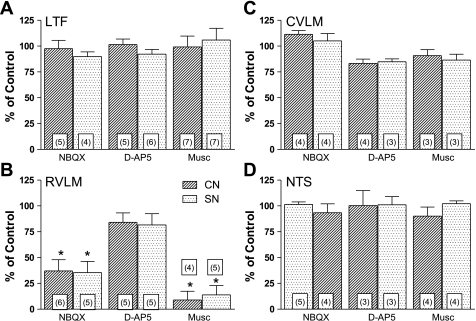
This study was designed to build on past work from this laboratory by testing the hypothesis that medullary lateral tegmental field (LTF) neurons play a critical role in mediating sympathoexcitatory responses to activation of sympathetic afferent fibers. We studied the effects of microinjection of N-methyl-d-aspartate (NMDA) or non-NMDA receptor antagonists or muscimol bilaterally into the LTF on the area under the curve of the computer-averaged sympathoexcitatory potential in the right inferior cardiac nerve elicited by short trains of stimuli applied to afferent fibers in the left inferior cardiac or left splanchnic nerve (CN, SN) of baroreceptor-denervated and vagotomized cats anesthetized with a mixture of diallylbarbiturate and urethane. In contrast to our hypothesis, sympathoexcitatory responses to stimulation of CN (n = 5-7) or SN (n = 4-7) afferent fibers were not significantly affected by these procedures. We then determined whether the rostral and caudal ventrolateral medulla (RVLM, CVLM) and nucleus tractus solitarius (NTS) were involved in mediating these reflexes. Blockade of non-NMDA, but not NMDA, receptors in the RVLM significantly reduced the area under the curve of the sympathoexcitatory responses to electrical stimulation of either CN (P = 0.0110; n = 6) or SN (P = 0.0131; n = 5) afferent fibers. Neither blockade of excitatory amino acid receptors nor chemical inactivation of CVLM or NTS significantly affected the responses. These data show that activation of non-NMDA receptors in the RVLM is a critical step in mediating the sympatho-sympathetic reflex.
Figures
Similar articles
-
Role of the medullary lateral tegmental field in reflex-mediated sympathoexcitation in cats.Am J Physiol Regul Integr Comp Physiol. 2004 Mar;286(3):R451-64. doi: 10.1152/ajpregu.00569.2003. Epub 2003 Nov 6. Am J Physiol Regul Integr Comp Physiol. 2004. PMID: 14604845
-
Involvement of non-NMDA and NMDA receptors in glutamate-induced pressor or depressor responses of the pons and medulla.Clin Exp Pharmacol Physiol. 1997 Jan;24(1):46-56. doi: 10.1111/j.1440-1681.1997.tb01782.x. Clin Exp Pharmacol Physiol. 1997. PMID: 9043805
-
Nucleus raphe pallidus participates in midbrain-medullary cardiovascular sympathoinhibition during electroacupuncture.Am J Physiol Regul Integr Comp Physiol. 2010 Nov;299(5):R1369-76. doi: 10.1152/ajpregu.00361.2010. Epub 2010 Aug 18. Am J Physiol Regul Integr Comp Physiol. 2010. PMID: 20720173 Free PMC article.
-
The role of the medullary lateral tegmental field in the generation and baroreceptor reflex control of sympathetic nerve discharge in the cat.Ann N Y Acad Sci. 2001 Jun;940:270-85. doi: 10.1111/j.1749-6632.2001.tb03683.x. Ann N Y Acad Sci. 2001. PMID: 11458684 Review.
-
Carotid chemoreflex. Neural pathways and transmitters.Adv Exp Med Biol. 1996;410:357-64. Adv Exp Med Biol. 1996. PMID: 9030325 Review.
Cited by
-
Does aging alter the molecular substrate of ionotropic neurotransmitter receptors in the rostral ventral lateral medulla? - A short communication.Exp Gerontol. 2017 May;91:99-103. doi: 10.1016/j.exger.2017.03.001. Epub 2017 Mar 2. Exp Gerontol. 2017. PMID: 28263869 Free PMC article.
-
A trigeminoreticular pathway: implications in pain.PLoS One. 2011;6(9):e24499. doi: 10.1371/journal.pone.0024499. Epub 2011 Sep 21. PLoS One. 2011. PMID: 21957454 Free PMC article.
-
2019 Ludwig Lecture: Rhythms in sympathetic nerve activity are a key to understanding neural control of the cardiovascular system.Am J Physiol Regul Integr Comp Physiol. 2020 Feb 1;318(2):R191-R205. doi: 10.1152/ajpregu.00298.2019. Epub 2019 Oct 30. Am J Physiol Regul Integr Comp Physiol. 2020. PMID: 31664868 Free PMC article. Review.
-
Responses of neurons in the caudal medullary lateral tegmental field to visceral inputs and vestibular stimulation in vertical planes.Am J Physiol Regul Integr Comp Physiol. 2012 Nov 1;303(9):R929-40. doi: 10.1152/ajpregu.00356.2012. Epub 2012 Sep 5. Am J Physiol Regul Integr Comp Physiol. 2012. PMID: 22955058 Free PMC article.
-
The Role of Angiotensin II Infusion on the Baroreflex Sensitivity and Renal Function in Intact and Bilateral Renal Denervation Rats.Adv Biomed Res. 2018 Mar 27;7:52. doi: 10.4103/abr.abr_192_17. eCollection 2018. Adv Biomed Res. 2018. PMID: 29657937 Free PMC article.
References
-
- Ammons WS, Foreman RD. Cardiovascular and T2-T4 dorsal horn cell responses to gallbladder distention in the cat. Brain Res 321: 267–277, 1984 - PubMed
-
- Barber WD, Yuan CS. Gastric vagal-splanchnic interactions in the brainstem of the cat. Brain Res 487: 1–8, 1989 - PubMed
-
- Barman SM, Gebber GL. Axonal projection patterns of ventrolateral medullospinal sympathoexcitatory neurons. J Neurophysiol 53: 1551–1566, 1985 - PubMed
-
- Barman SM, Gebber GL. Lateral tegmental field neurons of cat medulla: a source of basal activity of ventrolateral medullospinal sympathoexcitatory neurons. J Neurophysiol 57: 1410–1424, 1987 - PubMed
-
- Barman SM, Gebber GL. Lateral tegmental field neurons play a permissive role in governing the 10-Hz rhythm in sympathetic nerve discharge. Am J Physiol Regul Integr Comp Physiol 265: R1006–R1013, 1993 - PubMed
Publication types
MeSH terms
Substances
Grants and funding
LinkOut - more resources
Full Text Sources
Miscellaneous