

Innervation of the mouse cornea during development
- PMID: 20811061
- PMCID: PMC3053279
- DOI: 10.1167/iovs.10-5902
Innervation of the mouse cornea during development
Abstract
Purpose: Dense innervation of the cornea is important for maintaining its homeostasis and transparency. Although corneal nerves have been well studied in adults, little is known about mammalian corneal innervation during development. This study provides a detailed profile of nerves at various stages of mouse cornea development.
Methods: Mouse heads and corneas were collected at various stages of development including embryonic days (E)12.5 to E16.5, postnatal days (P)0, P10, three weeks after birth, and the adult. Corneas were immunostained with an anti-neuron-specific β-tubulin antibody (TUJ1). Fluorescently labeled nerves in whole-mount tissues and sections were imaged and analyzed for their axonal projections during eye development.
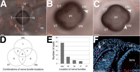
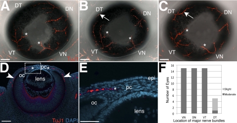
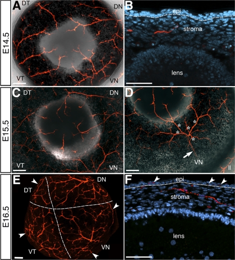
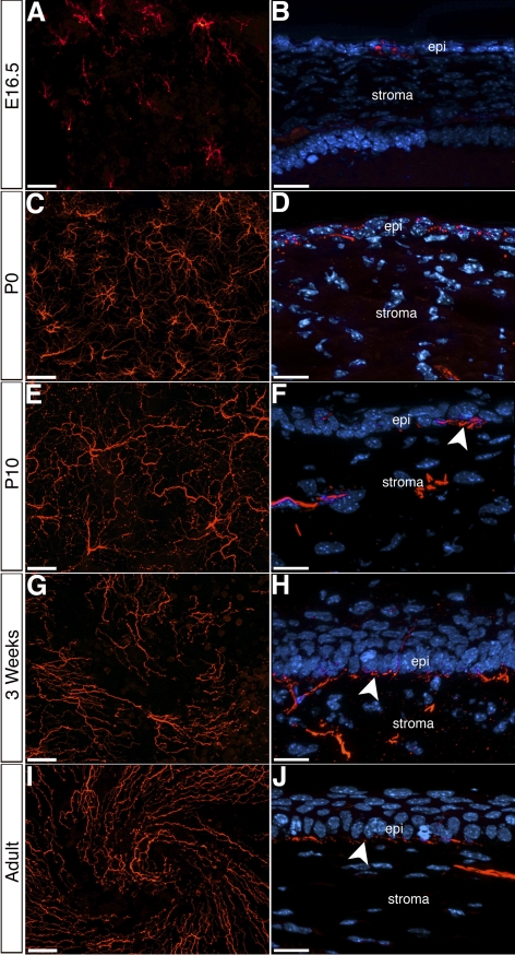
Results: The first nerve bundles appear at the periphery of the anterior portion of the eye by E12.5. Initial projection into the stroma occurs at E13.5 without formation of a pericorneal nerve ring. Between E13.5 and E16.5, nerve bundles project directly into the periphery of the presumptive cornea stroma. They branch repeatedly as they extend toward the cornea center and epithelium. Concomitantly, nerve bundles originating from four quadrants of the eye bifurcate into smaller branches that innervate the entire stroma. The first epithelial innervation occurs at E16.5. Epithelial nerves arrange into patterns that project toward the center subsequently forming a swirl at three weeks after birth, which becomes more pronounced in adults.
Conclusions: Nerve bundles that arise from four quadrants of the eye innervate the mouse cornea. The nerve bundles directly innervate the stroma without forming a pericorneal nerve ring. Radial arrangement of epithelial nerves gradually becomes centrally oriented, subsequently forming a swirl pattern.
Figures
Similar articles
-
Anatomy of the human corneal innervation.Exp Eye Res. 2010 Apr;90(4):478-92. doi: 10.1016/j.exer.2009.12.010. Epub 2009 Dec 29. Exp Eye Res. 2010. PMID: 20036654
-
Specificity of corneal nerve positions during embryogenesis.Mol Vis. 2001 Dec 21;7:297-304. Mol Vis. 2001. PMID: 11754335
-
[Embryogenesis and structural-and-functional specificity of corneal nerves in health and pathology].Vestn Oftalmol. 2004 Jul-Aug;120(4):47-50. Vestn Oftalmol. 2004. PMID: 15384854 Review. Russian. No abstract available.
-
Mapping the entire human corneal nerve architecture.Exp Eye Res. 2010 Oct;91(4):513-23. doi: 10.1016/j.exer.2010.07.007. Epub 2010 Jul 27. Exp Eye Res. 2010. PMID: 20650270 Free PMC article.
-
Corneal Development: Different Cells from a Common Progenitor.Prog Mol Biol Transl Sci. 2015;134:43-59. doi: 10.1016/bs.pmbts.2015.04.003. Epub 2015 Jun 4. Prog Mol Biol Transl Sci. 2015. PMID: 26310148 Review.
Cited by
-
The Clinical Guiding Role of the Distribution of Corneal Nerves in the Selection of Incision for Penetrating Corneal Surgery in Canines.Vet Sci. 2021 Dec 8;8(12):313. doi: 10.3390/vetsci8120313. Vet Sci. 2021. PMID: 34941840 Free PMC article.
-
Primary cilia deficiency in neural crest cells models anterior segment dysgenesis in mouse.Elife. 2019 Dec 17;8:e52423. doi: 10.7554/eLife.52423. Elife. 2019. PMID: 31845891 Free PMC article.
-
The miR-183/96/182 cluster is a checkpoint for resident immune cells and shapes the cellular landscape of the cornea.Ocul Surf. 2023 Oct;30:17-41. doi: 10.1016/j.jtos.2023.07.012. Epub 2023 Aug 2. Ocul Surf. 2023. PMID: 37536656 Free PMC article.
-
Regenerated Corneal Epithelium Expresses More βIII-Tubulin After Chemical Injuries Compared to Mechanical Injuries.Transl Vis Sci Technol. 2023 Dec 1;12(12):12. doi: 10.1167/tvst.12.12.12. Transl Vis Sci Technol. 2023. PMID: 38085248 Free PMC article.
-
Corneal epithelial development and homeostasis.Differentiation. 2023 Jul-Aug;132:4-14. doi: 10.1016/j.diff.2023.02.002. Epub 2023 Mar 1. Differentiation. 2023. PMID: 36870804 Free PMC article. Review.
References
-
- Marfurt CF, Kingsley RE, Echtenkamp SE. Sensory and sympathetic innervation of the mammalian cornea. A retrograde tracing study. Invest Ophthalmol Vis Sci. 1989;30:461–472 - PubMed
-
- Lwigale PY. Embryonic origin of avian corneal sensory nerves. Dev Biol. 2001;239:323–337 - PubMed
-
- Marfurt CF, Jones MA, Thrasher K. Parasympathetic innervation of the rat cornea. Exp Eye Res. 1998;66:437–448 - PubMed
-
- Morgan C, DeGroat WC, Jannetta PJ. Sympathetic innervation of the cornea from the superior cervical ganglion. An HRP study in the cat. J Auton Nerv Syst. 1987;20:179–183 - PubMed
-
- Tervo T. Demonstration of adrenergic nerve fibres in the nasociliary but not the ophthalmic nerve of the rat. Exp Eye Res. 1978;27:607–613 - PubMed
Publication types
MeSH terms
Substances
Grants and funding
LinkOut - more resources
Full Text Sources
