

Age-severity relationships in families linked to FCD2 with retroillumination photography
- PMID: 20811064
- PMCID: PMC3055756
- DOI: 10.1167/iovs.10-5187
Age-severity relationships in families linked to FCD2 with retroillumination photography
Abstract
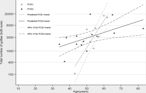
Purpose: Fuchs corneal dystrophy (FCD) is a progressive disorder of the corneal endothelium and is pathologically defined by the presence of guttae, which are excrescences of the Descemet membrane. The present study was undertaken to investigate the age-severity relationship of the FCD2-linked disease phenotype using retroillumination photography and to compare it with the characteristics of FCD1.
Methods: Two large families with multiple affected members were recruited. Exclusion analyses of the known late-onset FCD loci were completed with closely spaced STR markers, whereas genes associated with early- and late-onset FCD were investigated by bidirectional sequencing. Haplotypes were constructed, and two-point LOD scores were calculated. To document age-severity relationships, retroillumination photographs were acquired from members of both families.
Results: Parametric linkage and haplotype analysis mapped both families to FCD2 with significant two-point LOD scores. A total of 70,249 guttae were counted in 14 persons from both families. A significant increase in guttae density in the inferotemporal region (P = 0.016) was observed, a pattern similarly observed in a family linked to FCD1. Similarly, FCD2-linked families display an exponential trend in severity with age, as was observed in a family linked to FCD1. Finally, comparison of FCD1 and FCD2 exponential models suggested that the FCD1 phenotype is significantly more severe (P = 0.01).
Conclusions: A combination of genetic mapping and retroillumination photography was used to quantify the severity of the disease phenotype associated with FCD2 and to compare it to the disease characteristics of FCD1. These data suggest that this approach might have sufficient resolution to discriminate between discrete genetic FCD backgrounds, which will potentially aid in patient management.
Figures

References
-
- Lorenzetti DW, Uotila MH, Parikh N, Kaufman HE. Central cornea guttata: incidence in the general population. Am J Ophthalmol. 1967;64:1155–1158 - PubMed
-
- Krachmer JH, Purcell JJ, Jr, Young CW, Bucher KD. Corneal endothelial dystrophy: a study of 64 families. Arch Ophthalmol. 1978;96:2036–2039 - PubMed
-
- Mannis MJ, Holland EJ, Beck RW, et al. Clinical profile and early surgical complications in the Cornea Donor Study. Cornea. 2006;25:164–170 - PubMed
-
- Bergmanson JP, Sheldon TM, Goosey JD. Fuchs' endothelial dystrophy: a fresh look at an aging disease. Ophthalmic Physiol Opt. 1999;19:210–222 - PubMed
-
- Vogt A. Weitere Ergebnisse der Spaltlampenmikroskopie des vordern Bulbusabschnittes. Arch Ophthalmol. 1921:63–113
Publication types
MeSH terms
Substances
Supplementary concepts
Grants and funding
LinkOut - more resources
Full Text Sources
Medical
