

Osteogenic potential of mandibular vs. long-bone marrow stromal cells
- PMID: 20811069
- PMCID: PMC3113466
- DOI: 10.1177/0022034510378427
Osteogenic potential of mandibular vs. long-bone marrow stromal cells
Abstract
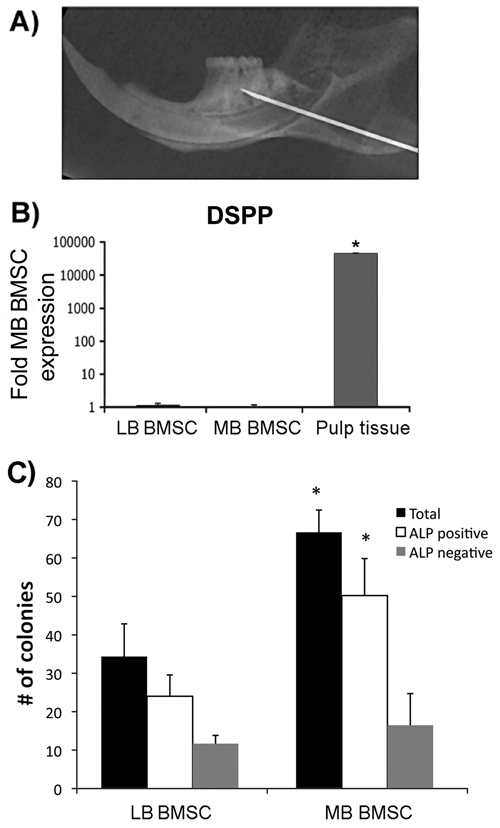
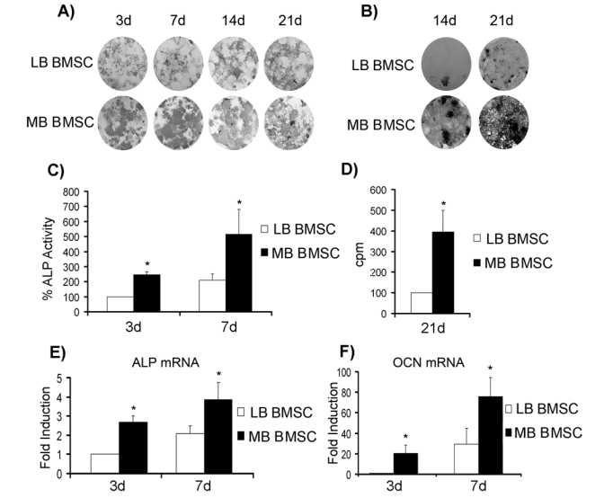
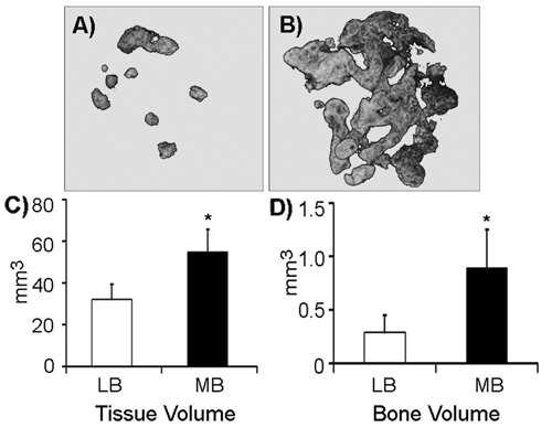
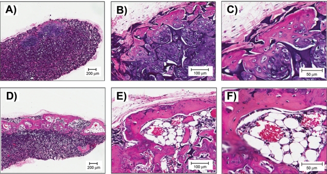
Although fundamentally similar to other bones, the jaws demonstrate discrete responses to developmental, mechanical, and homeostatic regulatory signals. Here, we hypothesized that rat mandible vs. long-bone marrow-derived cells possess different osteogenic potential. We established a protocol for rat mandible and long-bone marrow stromal cell (BMSC) isolation and culture. Mandible BMSC cultures formed more colonies, suggesting an increased CFU-F population. Both mandible and long-bone BMSCs differentiated into osteoblasts. However, mandible BMSCs demonstrated augmented alkaline phosphatase activity, mineralization, and osteoblast gene expression. Importantly, upon implantation into nude mice, mandible BMSCs formed 70% larger bone nodules containing three-fold more mineralized bone compared with long-bone BMSCs. Analysis of these data demonstrates an increased osteogenic potential and augmented capacity of mandible BMSCs to induce bone formation in vitro and in vivo. Our findings support differences in the mechanisms underlying mandible homeostasis and the pathophysiology of diseases unique to the jaws.
Figures
References
-
- Abzhanov A, Rodda SJ, McMahon AP, Tabin CJ. (2007). Regulation of skeletogenic differentiation in cranial dermal bone. Development 134:3133-3144 - PubMed
-
- Akintoye SO, Lam T, Shi S, Brahim J, Collins MT, Robey PG. (2006). Skeletal site-specific characterization of orofacial and iliac crest human bone marrow stromal cells in same individuals. Bone 38:758-768 - PubMed
-
- Chai Y, Maxson RE., Jr (2006). Recent advances in craniofacial morphogenesis. Dev Dyn 235:2353-2375 - PubMed
-
- Chai Y, Jiang X, Ito Y, Bringas P, Jr, Han J, Rowitch DH, et al. (2000). Fate of the mammalian cranial neural crest during tooth and mandibular morphogenesis. Development 127:1671-1679 - PubMed
-
- Daegling DJ, Hylander WL. (1997). Occlusal forces and mandibular bone strain: is the primate jaw “overdesigned”? J Hum Evol 33:705-717 - PubMed
Publication types
MeSH terms
Substances
Grants and funding
LinkOut - more resources
Full Text Sources
