. 2010 Oct;120(10):3480-92.
doi: 10.1172/JCI43165. Epub 2010 Sep 1.

Inhibition of TRPC6 degradation suppresses ischemic brain damage in rats

Affiliations

Inhibition of TRPC6 degradation suppresses ischemic brain damage in rats

Wanlu Du et al. J Clin Invest. 2010 Oct.

Abstract

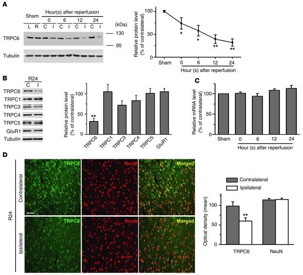
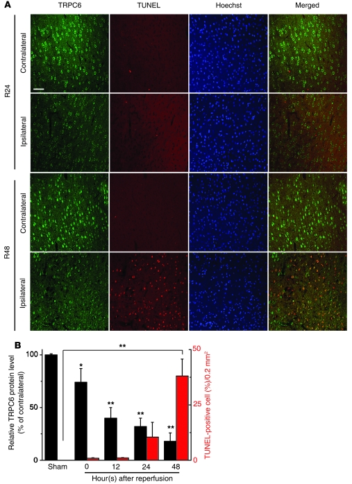
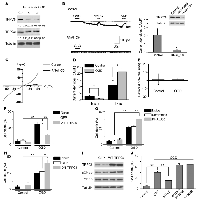
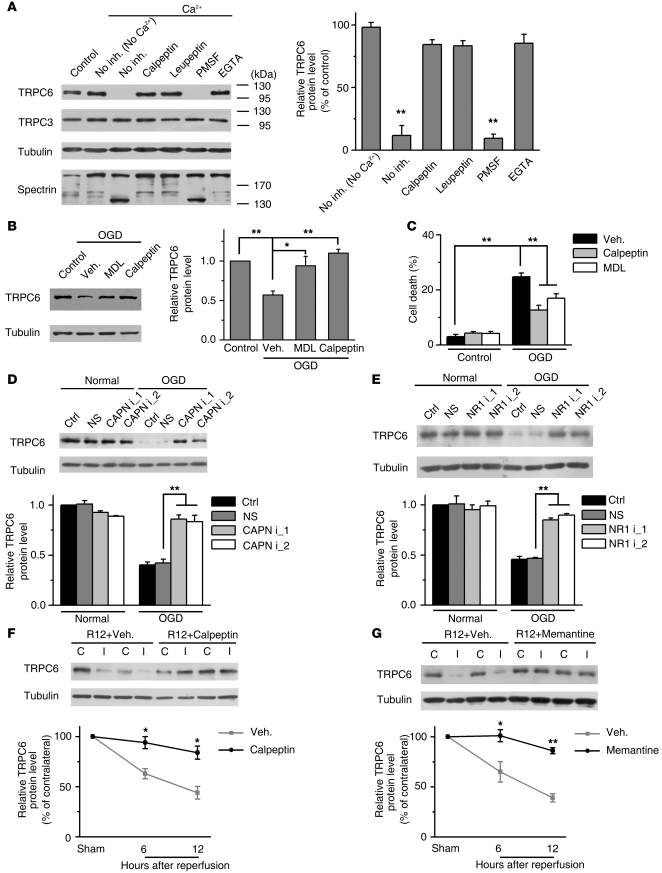
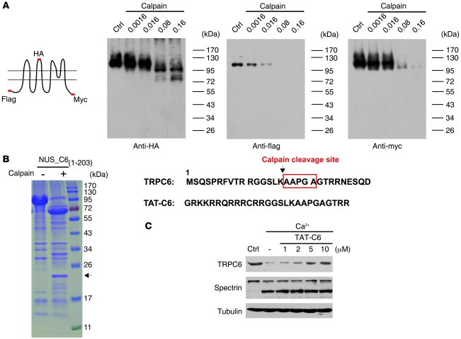
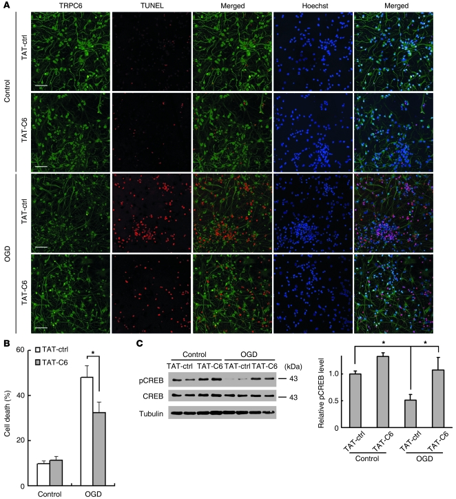
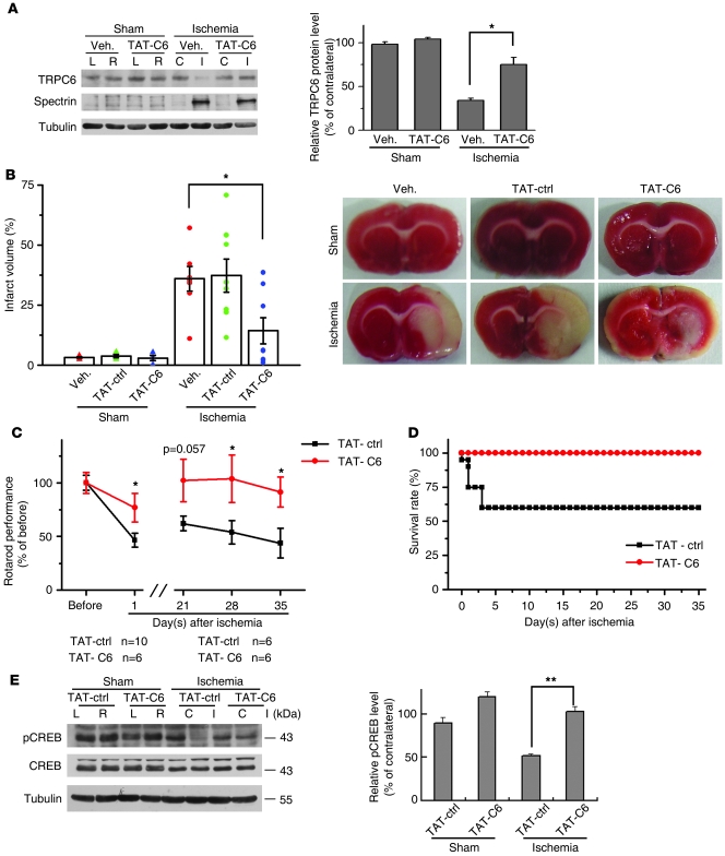
Brain injury after focal cerebral ischemia, the most common cause of stroke, develops from a series of pathological processes, including excitotoxicity, inflammation, and apoptosis. While NMDA receptors have been implicated in excitotoxicity, attempts to prevent ischemic brain damage by blocking NMDA receptors have been disappointing. Disruption of neuroprotective pathways may be another avenue responsible for ischemic damage, and thus preservation of neuronal survival may be important for prevention of ischemic brain injury. Here, we report that suppression of proteolytic degradation of transient receptor potential canonical 6 (TRPC6) prevented ischemic neuronal cell death in a rat model of stroke. The TRPC6 protein level in neurons was greatly reduced in ischemia via NMDA receptor-dependent calpain proteolysis of the N-terminal domain of TRPC6 at Lys¹⁶. This downregulation was specific for TRPC6 and preceded neuronal death. In a rat model of ischemia, activating TRPC6 prevented neuronal death, while blocking TRPC6 increased sensitivity to ischemia. A fusion peptide derived from the calpain cleavage site in TRPC6 inhibited degradation of TRPC6, reduced infarct size, and improved behavioral performance measures via the cAMP response element-binding protein (CREB) signaling pathway. Thus, TRPC6 proteolysis contributed to ischemic neuronal cell death, and suppression of its degradation preserved neuronal survival and prevented ischemic brain damage.


Figures

Figure 1
Figure 1. TRPC6 in the neurons was downregulated in ischemia.
(A) Immunoblots of the extracts from the contralateral (C) or ipsilateral (I) cortex (sham; left [L] or right [R] hemisphere) using the indicated antibodies. Tubulin served as a loading control. Right panel: quantification of the normalized TRPC6 protein levels (n = 5–8 rats per time point). *P < 0.05; **P < 0.01 versus sham. (B) Immunoblots for TRPCs and GluR1 after 24 hours reperfusion (R24). Right panel: quantification of the protein levels (n = 5 rats). **P < 0.01 versus contralateral. (C) TRPC6 mRNA levels determined by qRT-PCR (n = 3–5 rats per time point). (D) Representative images of the indicated cortex after 24 hours reperfusion (R24) double-stained with the indicated antibodies. Scale bar: 50 μm. Quantification of the optical density for both TRPC6 and NeuN immunoreactivity is shown on the right panel (n = 5 rats). **P < 0.01 versus the density in contralateral cortex. Data are presented as mean ± SEM. Unless stated, TRPC6 antibody from Millipore was used for Figures 1, 2, and 6 and antibody from Alomone Labs was used for Figures 3, 4, 5, and 7.
Figure 2
Figure 2. Downregulation of TRPC6 was prior to and during ischemic neuronal death.
(A) Representative images of the indicated cortex after 24 hours (R24) or 48 hours (R48) reperfusion double stained with TRPC6 antibody (Millipore) and TUNEL labeling. Hoechst stain was used to label the nucleus. Scale bar: 50 μm. (B) Quantification of the levels of TRPC6 and the numbers of TUNEL-positive cells in the ipsilateral cortex at the indicated times or sham surgery (sham) (n = 3 rats for each condition). *P < 0.05; **P < 0.01 versus sham. Data are presented as mean ± SEM.
Figure 3
Figure 3. TRPC6 was downregulated in modeled ischemia.
(A) Immunoblots of the extracts from cultured cortical neurons after OGD with indicated antibodies. The numbers below the blots are the normalized protein levels (n = 3). (B) Whole-cell recording for cultured cortical neurons (upper trace, control) or neurons transfected with RNAi_C6 (lower trace, RNAi_C6). Right panel: quantification of the current density. Islet: representative blots for TRPC6. OAG (100 μM); SKF, SKF96365 (10 μM). NMDG in Ca2+-free solution (n = 10 cells for control group; n = 8 cells for RNAi_C6 group). *P < 0.05 versus control. (C) I/V relationship of the OAG-induced currents from untransfected (control) or RNAi_C6-transfected neurons (RNAi_C6). Quantification of the indicated current densities (D) or the reversal potential of IOAG (E) from control or OGD-treated neurons. *P < 0.05 versus control. (FH) Quantification of cell death for the neurons transfected with the constructs, subjected to OGD followed by 24 hours incubation (n = 3). *P < 0.05; **P < 0.01 versus GFP or scrambled. (I) Immunoblots of the extracts from the neurons transfected with GFP or WT TRPC6 using the indicated antibodies. (J) Cortical neurons transfected with the constructs subjected to OGD and cell death assessed by PI staining. KCREB, dominant-negative form of CREB (n = 3). **P < 0.01 versus GFP. Data are presented as mean ± SEM.
Figure 4
Figure 4. NMDAR-mediated calpain proteolysis of TRPC6 in modeled ischemia.
(A) TRPC6 was proteolyzed by calpain. Immunoblots of the rat brain lysates incubated with Ca2+ in the presence or absence (No inh.) of the indicated agents using the indicated antibodies. Calpeptin, 20 μM; leupeptin, 100 μM; PMSF, 100 μM; EGTA, 5 mM. Right panel: quantification of TRPC6 levels (n = 3). **P < 0.01 versus control. (B) Immunoblots for TRPC6 in cortical neurons 24 hours after OGD in the presence of vehicle (Veh.), calpeptin (20 μM), or MDL28170 (MDL, 60 μM). Right panel: quantification of TRPC6 levels (n = 3). *P < 0.05; **P < 0.01 versus vehicle (TRPC6 antibody: Sigma-Aldrich). (C) Quantification of OGD-induced cell death (at 24 hours) in the presence of vehicle, calpeptin, or MDL28170 (n = 3). **P < 0.01 versus vehicle. (D) 2 calpain siRNA prevented OGD-induced TRPC6 degradation. Lower panel: quantification of TRPC6 levels (n = 3). **P < 0.01 versus nonsense siRNA (NS). (E) 2 NR1 siRNA inhibited OGD-induced TRPC6 degradation. Right panel: quantification of TRPC6 levels (n = 3) **P < 0.01 versus NS. Calpeptin (F) or memantine (G) suppressed the ischemia-induced TRPC6 degradation as analyzed by immunoblots. Quantifications of TRPC6 levels are shown on the lower panel. R12, 12 hours after reperfusion (n = 3). **P < 0.01 versus vehicle. Data are presented as mean ± SEM.
Figure 5
Figure 5. TRPC6 was cleaved by calpain at Lys16.
(A) Immunoblots of the lysates from HEK293 cells transfected with the TRPC6 tagged with N-flag-second loop-HA-C-myc (the schematic) and incubated with μ-calpain at the indicated concentrations using the indicated antibodies. (B) Purified NUS_C61-203 was digested with calpain and stained with Coomassie blue. Arrow indicates a 22-kDa fragment, whose N-terminal sequence is shown on the right. Lower sequence shows TAT-C6. (C) Immunoblots of the rat brain lysates incubated with 1 mM Ca2+ for 30 minutes in the presence of the indicated concentrations of TAT-C6 peptide using the indicated antibodies.
Figure 6
Figure 6. TAT-C6 inhibited neuronal cell death induced by OGD.
(A) Representative images of cortical neurons incubated with 5 μM TAT-ctrl or TAT-C6 2 hours before OGD and double stained with TRPC6 antibody and TUNEL labeling 24 hours after OGD. Hoechst stain was used to label the nucleus. Scale bars: 50 μm. (B) Quantification of OGD-induced cell death (at 24 hours) in the presence of TAT-ctrl or TAT-C6. *P < 0.05 versus TAT-ctrl. (C) Immunoblots for CREB and pCREB of the extracts from OGD-treated cortical neurons receiving TAT-ctrl or TAT-C6. Right panel shows quantification of p-CREB levels (n = 3–6 cultures). *P < 0.05 versus control or TAT-ctrl. Data are presented as mean ± SEM.
Figure 7
Figure 7. Inhibiting calpain-mediated TRPC6 degradation protected rat brains from ischemic damage.
(A) Immunoblots for TRPC6 and spectrin in the cortical extracts from ischemic or sham-treated rats receiving vehicle or TAT-C6 peptide (TAT-C6). Right panel: quantification of TRPC6 protein levels (n = 3 rats). *P < 0.05 versus vehicle. (B) TTC-stained brain sections indicated the infarct volumes (bar graph) or area (image) in rats receiving vehicle, control TAT-peptide (TAT-ctrl), or TAT-C6 peptide after ischemia (n = 8 rats). *P < 0.05 versus vehicle. (C) Rotarod evaluation of the 2 groups of rats, showing behavioral improvement. Upper curve, rats injected with TAT-C6; lower curve, rats injected with TAT-ctrl. *P < 0.05 versus TAT-ctrl (below, number of rats used for statistical analysis). (D) Survival rates of rats injected with TAT-C6 or TAT-ctrl (n = 6 for TAT-C6 group; n = 10 for TAT-ctrl group) after cerebral ischemia. (E) Immunoblots for CREB and pCREB in the cortical extracts from ischemic or sham-treated rats receiving TAT-ctrl or TAT-C6. Right panel: quantification of pCREB levels (n = 3 rats). **P < 0.01 versus TAT-ctrl. Data are presented as mean ± SEM.

Similar articles

Cited by

References

    1. Dirnagl U, Iadecola C, Moskowitz MA. Pathobiology of ischaemic stroke: an integrated view. Trends Neurosci. 1999;22(9):391–397. doi: 10.1016/S0166-2236(99)01401-0. - DOI - PubMed
    1. Lo EH, Dalkara T, Moskowitz MA. Mechanisms, challenges and opportunities in stroke. Nat Rev Neurosci. 2003;4(5):399–415. - PubMed
    1. Lipton P. Ischemic cell death in brain neurons. Physiol Rev. 1999;79(4):1431–1568. - PubMed
    1. Choi DW. Ischemia-induced neuronal apoptosis. Curr Opin Neurobiol. 1996;6(5):667–672. doi: 10.1016/S0959-4388(96)80101-2. - DOI - PubMed
    1. Hoyte L, Barber PA, Buchan AM, Hill MD. The rise and fall of NMDA antagonists for ischemic stroke. Curr Mol Med. 2004;4(2):131–136. - PubMed

Publication types

MeSH terms