. 2010 Oct;120(10):3520-9.
doi: 10.1172/JCI42028. Epub 2010 Sep 1.

Cardiac fibrosis in mice with hypertrophic cardiomyopathy is mediated by non-myocyte proliferation and requires Tgf-β

Affiliations

Cardiac fibrosis in mice with hypertrophic cardiomyopathy is mediated by non-myocyte proliferation and requires Tgf-β

Polakit Teekakirikul et al. J Clin Invest. 2010 Oct.

Abstract

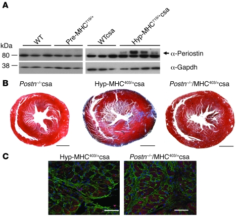
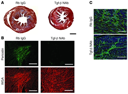
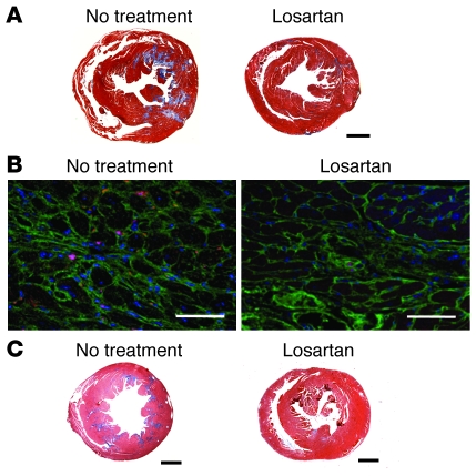
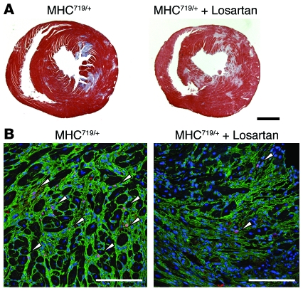
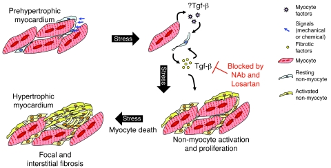
Mutations in sarcomere protein genes can cause hypertrophic cardiomyopathy (HCM), a disorder characterized by myocyte enlargement, fibrosis, and impaired ventricular relaxation. Here, we demonstrate that sarcomere protein gene mutations activate proliferative and profibrotic signals in non-myocyte cells to produce pathologic remodeling in HCM. Gene expression analyses of non-myocyte cells isolated from HCM mouse hearts showed increased levels of RNAs encoding cell-cycle proteins, Tgf-β, periostin, and other profibrotic proteins. Markedly increased BrdU labeling, Ki67 antigen expression, and periostin immunohistochemistry in the fibrotic regions of HCM hearts confirmed the transcriptional profiling data. Genetic ablation of periostin in HCM mice reduced but did not extinguish non-myocyte proliferation and fibrosis. In contrast, administration of Tgf-β-neutralizing antibodies abrogated non-myocyte proliferation and fibrosis. Chronic administration of the angiotensin II type 1 receptor antagonist losartan to mutation-positive, hypertrophy-negative (prehypertrophic) mice prevented the emergence of hypertrophy, non-myocyte proliferation, and fibrosis. Losartan treatment did not reverse pathologic remodeling of established HCM but did reduce non-myocyte proliferation. These data define non-myocyte activation of Tgf-β signaling as a pivotal mechanism for increased fibrosis in HCM and a potentially important factor contributing to diastolic dysfunction and heart failure. Preemptive pharmacologic inhibition of Tgf-β signals warrants study in human patients with sarcomere gene mutations.


Figures

Figure 1
Figure 1. Hypertrophic α-MHC719/+ mice have cardiac hypertrophy, increased myocardial fibrosis, and non-myocyte cell proliferation.
(A) Masson trichrome–stained sections from prehypertrophic (Pre-) and hypertrophic (Hyp-) α-MHC719/+csa hearts revealed increased fibrosis (blue) in comparison with WTcsa hearts. Scale bar: 1 mm. (B) Light microscopy shows increased BrdU incorporation in sections from α-MHC719/+csa hearts (top panel, original magnification, ×50). Confocal immunofluorescence after WGA (green), DAPI (blue) stains, and BrdU antibodies revealed proliferating non-myocyte cells (magenta nuclei, arrowheads) in regions with increased fibrosis (bottom panel, original magnification, ×400). There was minor nonspecific antibody staining (*) outside the nuclei. In comparison with WTcsa heart sections, note that α-MHC719/+csa hearts have disorganized myocardial architecture. Scale bar: 75 μm. (C) Confocal immunofluorescence shows expression of α-smooth muscle actin and fibroblast-specific protein 1 (Fsp1/S100a4), detected using a FITC-conjugated α-smooth muscle actin mouse antibody (green) and S100a4 antibody (red), respectively. Note that FITC-conjugated α-smooth muscle actin labeled only vascular beds in WTcsa and hypertrophic α-MHC719/+csa hearts. Scale bar: 75 μm.
Figure 2
Figure 2. Increased periostin protein in hypertrophic α-MHC403/+ and α-MHC719/+ mice.
(A) Western blots detected periostin protein (arrow) in LV extracts from WT, prehypertrophic α-MHC719/+, WTcsa, and hypertrophic α-MHC719/+csa mice but not in extracts from Postn–/– mice (data not shown). Gapdh served as a control for loading. Periostin levels were variable in hypertrophic α-MHC719/+csa mice, but densitometry indicated significant increases (P = 0.035) above levels in WTcsa mice. (B) Histopathological comparison of fibrotic regions in hearts from Postn–/–csa, hypertrophic α-MHC403/+csa, and Postn–/–α-MHC403/+csa mice. Scale bar: 1 mm. (C) Immunofluorescent images of LV sections stained for Ki67 antigen (magenta) and DAPI (blue) indicated proliferating cells in fibrotic areas (identified by WGA, green). Scale bar: 75 μm. Numbers of Ki67 antigen-positive nuclei in fibrotic regions of Postn–/–α-MHC403/+csa mice were significantly less than those in α-MHC403/+csa mice.
Figure 3
Figure 3. Tgf-β NAb attenuated cardiac fibrosis, reduced periostin expression, and diminished non-myocyte proliferation in hypertrophic α-MHC719/+csa mice.
(A) Histopathologic sections of Tgf-β NAb–treated and rabbit (Rb) IgG-treated α-MHC719/+csa mice stained with Masson trichrome (blue) demonstrated less fibrosis in Tgf-β NAb–treated mice. Scale bar: 1 mm. (B) Immunofluorescent images of LV sections stained for periostin (green) and WGA (red) showed reduced periostin expression in Tgf-β NAb–treated α-MHC719/+ mice and smaller areas of fibrosis compared with rabbit-IgG treated mice. Scale bar: 0.5 mm. (C) Confocal immunofluorescent images of LV sections from BrdU-treated mice, analyzed using BrdU antibodies (magenta), WGA (green), and DAPI (blue), showed that Tgf-β NAb treatment reduced the numbers of proliferating cells in fibrotic areas. Scale bar: 75 μm.
Figure 4
Figure 4. Losartan attenuated cardiac fibrosis, reduced periostin expression, and diminished non-myocyte proliferation in hypertrophic α-MHC719/+csa mice.
(A) Histopathologic sections stained with Masson trichrome stain (blue) revealed minimal fibrosis in losartan-treated α-MHC719/+csa compared with untreated α-MHC719/+ csa mice. Scale bar: 1 mm. (B) Confocal immunofluorescence images of LV sections stained for Ki67 antigen (magenta), WGA (green), and DAPI (blue) showed that losartan reduced non-myocyte proliferation. Scale bar: 60.6 μm. (C) Cardiac sections stained with Masson trichrome after chronic (30 weeks) losartan treatment in α-MHC719/+ mice showed markedly reduced amounts of myocardial fibrosis compared with untreated age-matched α-MHC719/+ mice. Note that these 35-week-old α-MHC719/+ mice did not receive CsA. Scale bar: 1 mm.
Figure 5
Figure 5. Losartan did not reverse fibrotic remodeling in established HCM but diminished non-myocyte proliferation.
(A) Cardiac sections from hypertrophic α-MHC719/+csa mice, treated or not treated with losartan, stained with Masson trichrome showed comparable amounts of myocardial fibrosis. Scale bar: 1 mm. (B) Non-myocyte proliferation, assessed by BrdU incorporation, was significantly reduced by losartan treatment compared with that of untreated mice in regions of fibrosis (WGA, green; DAPI, blue). Arrowheads indicate BrdU-positive nuclei. Scale bar: 75 μm.
Figure 6
Figure 6. A model for increasing fibrosis and diastolic dysfunction from HCM sarcomere gene mutations.
Mutant myocytes have increased biophysical properties (8) and abnormal Ca2+ homeostasis (11), factors that trigger mechanical and/or biochemical signals that activate gene transcription, including increased Tgf-β expression. Whether by paracrine and/or autocrine signaling, Tgf-β stimulates non-myocyte proliferation and expression of profibrotic molecules. Activated non-myocyte cells secrete profibrotic molecules that expand the interstitium, increase stresses imposed on mutant myocytes, and promote myocyte death with resultant focal scarring. Tgf-β–mediated increased interstitial and focal fibrosis contributes to diastolic dysfunction in HCM hearts. Preemptive antagonism of Tgf-β signaling by a neutralizing antibody (NAb) or losartan reduces non-myocyte proliferation and profibrotic gene expression, thereby limiting cardiac fibrosis.

References

    1. Seidman JG, Seidman C. The genetic basis for cardiomyopathy: from mutation identification to mechanistic paradigms. Cell. 2001;104(4):557–567. doi: 10.1016/S0092-8674(01)00242-2. - DOI - PubMed
    1. Maron BJ, Seidman JG, Seidman CE. Proposal for contemporary screening strategies in families with hypertrophic cardiomyopathy. J Am Coll Cardiol. 2004;44(11):2125–2132. doi: 10.1016/j.jacc.2004.08.052. - DOI - PubMed
    1. Maron BJ. Cardiology patient pages. Hypertrophic cardiomyopathy. Circulation. 2002;106(19):2419–2421. doi: 10.1161/01.CIR.0000034170.83171.0B. - DOI - PubMed
    1. Dhandapany PS, et al. A common MYBPC3 (cardiac myosin binding protein C) variant associated with cardiomyopathies in South Asia. Nat Genet. 2009;41(2):187–191. doi: 10.1038/ng.309. - DOI - PMC - PubMed
    1. Maron BJ. Hypertrophic cardiomyopathy in China. Am J Cardiol. 2007;100(1):145–146. doi: 10.1016/j.amjcard.2007.02.069. - DOI - PubMed

Publication types