Review
. 2010 Sep;120(9):3042-53.
doi: 10.1172/JCI42258. Epub 2010 Sep 1.

Lighting a candle in the dark: advances in genetics and gene therapy of recessive retinal dystrophies

Affiliations
Review

Lighting a candle in the dark: advances in genetics and gene therapy of recessive retinal dystrophies

Anneke I den Hollander et al. J Clin Invest. 2010 Sep.

Erratum in

  • J Clin Invest. 2011 Jan 4;121(1):456-7

Abstract

Nonsyndromic recessive retinal dystrophies cause severe visual impairment due to the death of photoreceptor and retinal pigment epithelium cells. These diseases until recently have been considered to be incurable. Molecular genetic studies in the last two decades have revealed the underlying molecular causes in approximately two-thirds of patients. The mammalian eye has been at the forefront of therapeutic trials based on gene augmentation in humans with an early-onset nonsyndromic recessive retinal dystrophy due to mutations in the retinal pigment epithelium-specific protein 65kDa (RPE65) gene. Tremendous challenges still lie ahead to extrapolate these studies to other retinal disease-causing genes, as human gene augmentation studies require testing in animal models for each individual gene and sufficiently large patient cohorts for clinical trials remain to be identified through cost-effective mutation screening protocols.


Figures
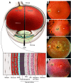
Figure 1
Figure 1. Anatomy of the human eye and retina.
(A) Cross section showing the major landmarks of the human eye and retina. The borders of the macula, which is adjacent to the optic disc, are indicated with a dashed line. Fundus views of patients with (B) normal vision; (C) retinitis pigmentosa (pigmentary changes indicated by arrowheads); (D) STGD1 due to ABCA4 mutations; and (E) LCA due to a homozygous RPE65 mutation. +, fovea; od, optic disc; INL inner nuclear layer; ONL, outer nuclear layer.
Figure 2
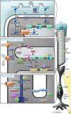
Figure 2. Schematic representation of three major processes in human rod photoreceptor cells and the RPE.
Upper panel: The retinoid cycle taking place in rod photoreceptor cells (PC) and the RPE. Upon photactivation, 11-cis-retinal is converted into all-trans-retinal and dissociates from activated rhodopsin. The all-trans-retinal is then recycled to produce more 11-cis-retinal via several enzymatic steps in the RPE. ABCA4 mediates transport of all-trans-retinal to the outside of the photoreceptor outer segment disks. The localization and function of proteins involved in AR and XL nonsyndromic retinal dystrophies are depicted, with the exception of GCAP, a critical Ca2+-binding interactor of GUCY2D, which is mutated in autosomal dominant CRD ( http://www.sph.uth.tmc.edu/Retnet/home.htm). CRALBP, protein product of RLBP1; IRBP, protein product of RBP3; RAL, retinal; RE, retinyl esters; RHOa, photoactivated rhodopsin; ROL, retinol. Middle panel: The phototransduction cascade in rod PCs. Upon photoactivation, amplification of the signal is mediated through the α-subunit of transducin and phosphodiesterase, which results in closure of the cGMP-gated channel, hyperpolarization of the cell, and reduced glutamate release at the synapse. SAG, arrestin. Lower panel: Ciliary transport along the connecting cilium. Kinesin II family motors mediate transport toward the outer segments; cytoplasmic dynein 2/1b (DYNC2H1) is involved in transport processes from the outer segments toward the inner segments. The precise roles of CEP290, Lebercilin, RPGR, RPGRIP1, and RP1 in ciliary transport processes are not yet known. AIPL1 (not indicated in this figure) is a chaperone for proteins that are farnesylated. For IDH3B and PRCD, the exact cellular functions are not known. ADAM9, MERTK, and RGR are secreted by the RPE and localize in the interphotoreceptor matrix. The CNGA3, CNGB3, GNAT2, and PDE6C genes are specifically expressed in cone PCs and therefore not indicated in this figure. At the right side, a Müller cell (MC) connects to the photoreceptor cell with the transmembrane protein CRB1. Usherin, protein product of USH2A.
Figure 3
Figure 3. Phenotypic overlap among autosomal recessive retinal dystrophies.
Patients with ACHM display a virtually stationary cone defect in which cones are principally defective. At end stage, CD can hardly be distinguished from CRD. Patients with STGD1 later in life show mid-peripheral defects similar to those in CRD patients. Patients with RP initially display tunnel vision due to rod defects that very often progresses to complete blindness when the cones are also affected. In patients with LCA, the defects can occur in both types of photoreceptors, or in RPE cells, and therefore clinical and molecular genetic overlap with CD, CRD, or RP can be expected.
Figure 4
Figure 4. Prevalence of mutations in genes causing genetically heterogeneous retinal dystrophies.
Estimated prevalence of mutations in genes causing AR RP, LCA, AR CRD, AR CD, and ACHM. Mutations in approximately 50% of AR RP, 30% of LCA, 60% of AR CRD, 90% of AR CD, and 20% of ACHM remain to be identified. For several genes, only one or a few families with mutations have been reported; in these cases, the gene frequency was estimated to be 1%. Estimates are based on literature searches ( http://www.sph.uth.tmc.edu/Retnet/home.htm) and our own experience.
Figure 5
Figure 5. Approaches for surgical delivery of gene therapy vectors in retinal disease.
Subretinal injection is necessary to place the gene augmentation therapy reagent in contact with the target photoreceptor and RPE cells. Arrows indicate the approaches used in the postnatal/adult mouse (A), large animals/humans (B), and fetal mice (C; injection into the subretinal space adjacent to retinal progenitor cells). (DG) Frames from an intraoperative video taken during subretinal injection of the macula in a human with rAAV2.hRPE65v2 (64). In G, the cannula is withdrawn, revealing the raised fovea (black arrowhead). *optic disc; white arrowheads indicate edge of the expanding detachment.

References

    1. Heckenlively J.Retinitis Pigmentosa . New York, New York, USA: Lippincott Williams & Wilkins; 1988.
    1. den Hollander AI, Roepman R, Koenekoop RK, Cremers FP. Leber congenital amaurosis: genes, proteins and disease mechanisms. Prog Retin Eye Res. 2008;27(4):391–419. doi: 10.1016/j.preteyeres.2008.05.003. - DOI - PubMed
    1. Blacharski PA. Fundus flavimaculatus. In: Newsome DA, ed.Retinal Dystrophies And Degenerations . New York, New York, USA: Raven Press; 1988.
    1. Walia S, Fishman GA. Natural history of phenotypic changes in Stargardt macular dystrophy. Ophthalmic Genet. 2009;30(2):63–68. doi: 10.1080/13816810802695550. - DOI - PubMed
    1. Michaelides M, Hunt DM, Moore AT. The cone dysfunction syndromes. Br J Ophthalmol. 2004;88(2):291–297. doi: 10.1136/bjo.2003.027102. - DOI - PMC - PubMed

Publication types

MeSH terms