Review
. 2010 Sep;120(9):3073-83.
doi: 10.1172/JCI42440. Epub 2010 Sep 1.

A look at autoimmunity and inflammation in the eye

Affiliations
Review

A look at autoimmunity and inflammation in the eye

Rachel R Caspi. J Clin Invest. 2010 Sep.

Abstract

Autoimmune and inflammatory uveitis are a group of potentially blinding intraocular inflammatory diseases that arise without a known infectious trigger and are often associated with immunological responses to unique retinal proteins. In the United States, about 10% of the cases of severe visual handicap are attributed to this group of disorders. As I discuss here, experimental models of ocular autoimmunity targeting retinal proteins have brought about a better understanding of the basic immunological mechanisms involved in the pathogenesis of uveitis and are serving as templates for the development of novel therapies.


Figures

Figure 1
Figure 1. Scheme of an eye showing major anatomical structures and organization.
Detail: Enlarged section of retina and uvea showing anatomical layers. Light passes through the ocular media and concentrates on the macula (the area of the retina responsible for sharp color vision), whereupon the photoreceptor cells sense the signal and transmit it to the brain via the optic nerve. Any damage to the ocular structures along the visual axis would likely result in a visual deficit. RPE, retinal pigment epithelium.
Figure 2
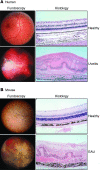
Figure 2. Clinical and histological appearance of uveitis: human and mouse.
(A) Appearance of the fundus (posterior pole of the eye) and retinal histology (original magnification, ×200) of a healthy human retina and those from a patient with uveitis. Images provided by Chi-Chao Chan, National Eye Institute, NIH. (B) Fundus (×20) and retinal histology (×400) of a B10.RIII mouse immunized with IRBP. Lower-right panels of A and B reproduced with permission from Immunological Reviews (24).
Figure 3
Figure 3. Critical checkpoints in uveitis, as defined from studies with animal models.
Retinal antigen–specific T cells that have not been eliminated in the thymus encounter an activating stimulus in the context of costimulatory “danger” signals, escape from the control of nTregs, and differentiate into pathogenic effector T cells. These undergo clonal expansion, migrate to the eye, break down the blood-retinal barrier, and recruit inflammatory leukocytes from the circulation. The resulting inflammation results in damage to the tissue and release of ocular antigens, which triggers eye-specific regulatory mechanisms that terminate the disease and limit pathology. mTECs, medullary thymic epithelial cells. Modified with permission from Immunological Reviews (24).

References

    1. Gery I, Nussenblatt RB, Chan CC, Caspi RR. Autoimmune diseases of the eye. In: Theofilopoulos AN, Bona CA, eds.The Molecular Pathology of Autoimmune Diseases . New York, New York, USA: Taylor and Francis; 2002:978–998.
    1. Gritz DC, Wong IG. Incidence and prevalence of uveitis in Northern California; the Northern California Epidemiology of Uveitis Study. Ophthalmology. 2004;111(3):491–500. doi: 10.1016/j.ophtha.2003.06.014. - DOI - PubMed
    1. Nussenblatt RB, Whitcup SM.Uveitis: Fundamentals and Clinical Practice . Philadelphia, Pennsylvania, USA: Mosby Publishing; 2004.
    1. Gocho K, Kondo I, Yamaki K. Identification of autoreactive T cells in Vogt-Koyanagi-Harada disease. Invest Ophthalmol Vis Sci. 2001;42(9):2004–2009. - PubMed
    1. Gery I, Mochizuki M, Nussenblatt RB. Retinal specific antigens and immunopathogenic processes they provoke. Prog Retinal Res. 1986;5:75–109.