. 2010 Sep;126(3):121e-133e.
doi: 10.1097/PRS.0b013e3181e605be.

Treatment of common congenital hand conditions

Affiliations

Treatment of common congenital hand conditions

Takashi Oda et al. Plast Reconstr Surg. 2010 Sep.

Abstract

Learning objectives: After reading this article, the participant should be able to: 1. Recognize the clinical features associated with five common congenital hand conditions. 2. Describe the indications and appropriate timing for various surgical procedures used to treat congenital hand anomalies. 3. Identify the pearls and pitfalls of these surgical treatments to avoid complications. 4. Understand the expected postoperative outcomes associated with these surgical procedures.

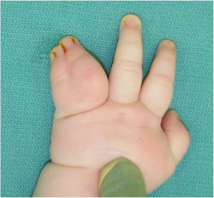
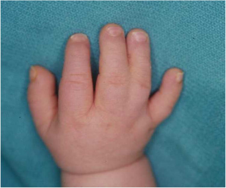
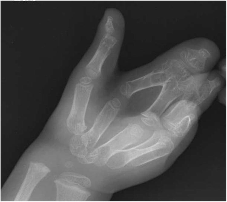
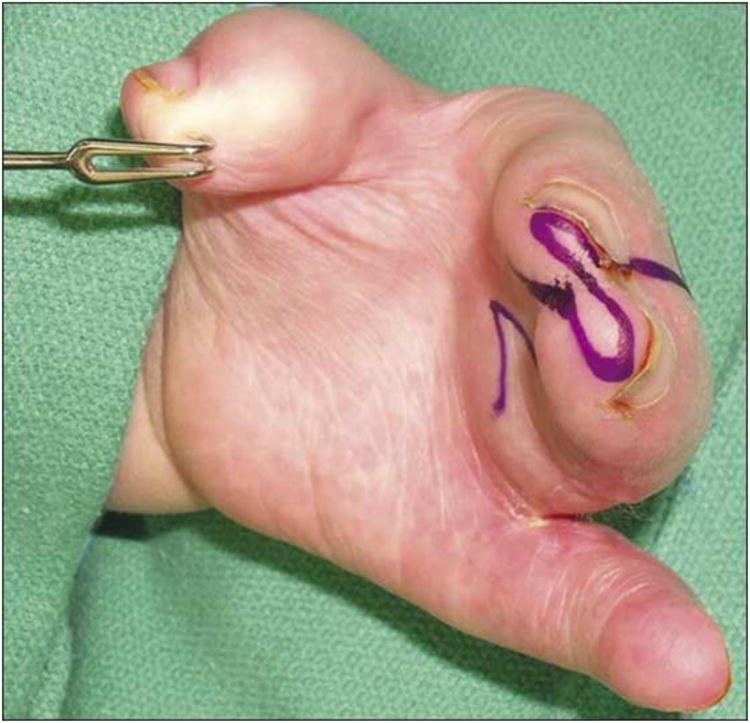
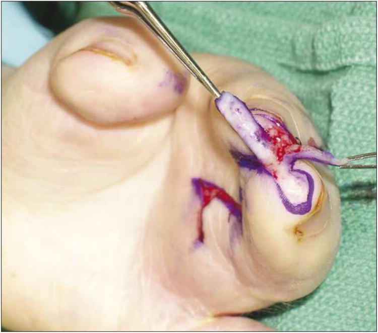
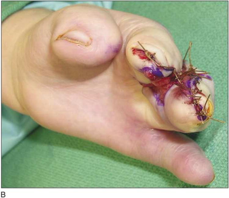
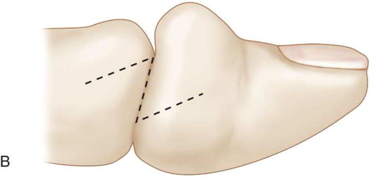
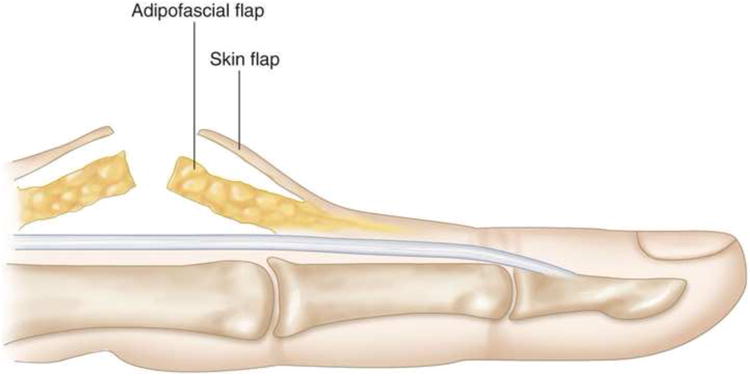
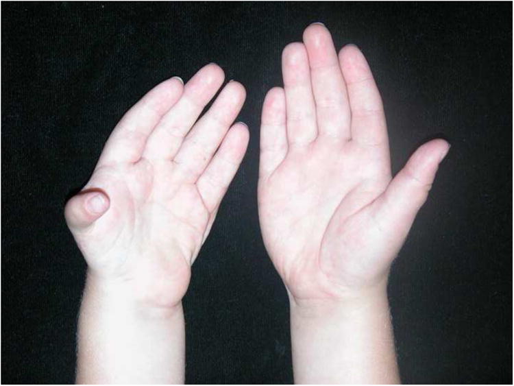
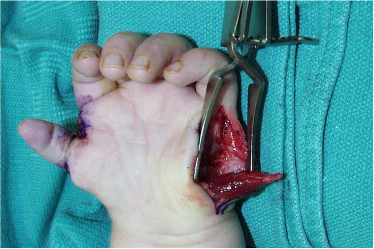
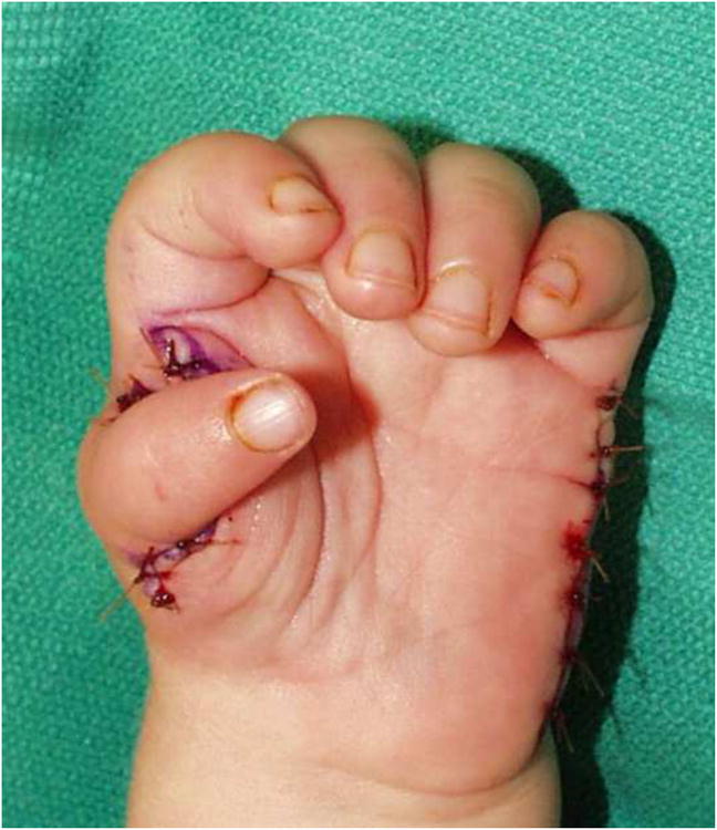
Summary: This article provides an introduction to congenital hand differences by focusing on practical surgical strategies for treating five commonly encountered conditions, including syndactyly, constriction ring syndrome, duplicated thumb, hypoplastic thumb, and trigger thumb. The accompanying videos demonstrate common and reliable surgical techniques for syndactyly release, duplicated thumb reconstruction, and pollicization for hypoplastic thumb.


Figures

Figure 1
Figure 1
Syndactyly. (A) Complete and simple syndactyly of the left ring and little fingers. (B) Incomplete and simple syndactyly of the right middle and ring fingers. (C) Complex syndactyly of the left middle and ring fingers. (D) The radiograph of the case (C).
Figure 1
Figure 1
Syndactyly. (A) Complete and simple syndactyly of the left ring and little fingers. (B) Incomplete and simple syndactyly of the right middle and ring fingers. (C) Complex syndactyly of the left middle and ring fingers. (D) The radiograph of the case (C).
Figure 1
Figure 1
Syndactyly. (A) Complete and simple syndactyly of the left ring and little fingers. (B) Incomplete and simple syndactyly of the right middle and ring fingers. (C) Complex syndactyly of the left middle and ring fingers. (D) The radiograph of the case (C).
Figure 1
Figure 1
Syndactyly. (A) Complete and simple syndactyly of the left ring and little fingers. (B) Incomplete and simple syndactyly of the right middle and ring fingers. (C) Complex syndactyly of the left middle and ring fingers. (D) The radiograph of the case (C).
Figure 2
Figure 2
Nail fold reconstruction. (A) Skin flaps are designed on the tip of digits. (B) The skin flaps are elevated before separation of digits. (C) Each skin flap comes from the adjacent digit and is sutured onto the nail. (Reprinted with permission from Kim, S. E., Chung, K. C. Syndactyly release. In K. C. Chung (Ed.), Operative technique; hand and wrist surgery, Vol. 2. Philadelphia: Saunders Elsevier, 2008. Pp. 847-858.)
Figure 2
Figure 2
Nail fold reconstruction. (A) Skin flaps are designed on the tip of digits. (B) The skin flaps are elevated before separation of digits. (C) Each skin flap comes from the adjacent digit and is sutured onto the nail. (Reprinted with permission from Kim, S. E., Chung, K. C. Syndactyly release. In K. C. Chung (Ed.), Operative technique; hand and wrist surgery, Vol. 2. Philadelphia: Saunders Elsevier, 2008. Pp. 847-858.)
Figure 3
Figure 3
Z-plasty design on the lateral side of the digit. (Reprinted with permission from Chung, K. C., Kim, S. E. Correction of constriction ring. In K. C. Chung (Ed.), Operative technique; hand and wrist surgery, Vol. 2. Philadelphia: Saunders Elsevier, 2008. Pp. 837-846.)
Figure 4
Figure 4
Advancement of adipose flap. (A) Proximal and distal adipose flaps are elevated from paratenon. (B) Both adipose flaps are advanced into the defect of subcutaneous tissue. (Reprinted with permission from Chung, K. C., Kim, S. E. Correction of constriction ring. In K. C. Chung (Ed.), Operative technique; hand and wrist surgery, Vol. 2. Philadelphia: Saunders Elsevier, 2008. Pp. 837-846.)
Figure 4
Figure 4
Advancement of adipose flap. (A) Proximal and distal adipose flaps are elevated from paratenon. (B) Both adipose flaps are advanced into the defect of subcutaneous tissue. (Reprinted with permission from Chung, K. C., Kim, S. E. Correction of constriction ring. In K. C. Chung (Ed.), Operative technique; hand and wrist surgery, Vol. 2. Philadelphia: Saunders Elsevier, 2008. Pp. 837-846.)
Figure 5
Figure 5
Blood supply to the finger. Perforators from digital arteries supply the skin of the finger. Venus return is maintained by venae comitantes around the digital arteries. (Reprinted with permission from Chung, K. C., Kim, S. E. Correction of constriction ring. In K. C. Chung (Ed.), Operative technique; hand and wrist surgery, Vol. 2. Philadelphia: Saunders Elsevier, 2008. Pp. 837-846.)
Figure 6
Figure 6
Illustration of the modified Bilhaut-Cloquet procedure. Black portion is excised in order to preserve the IP joint. (A) For Wassel classification Type II. (B) For Wassel classification Type III. (Modified from Baek, G. H., Gong, H. S., Chung, M. S., et al. Modified Bilhaut-Cloquet procedure for Wassel type-II and III polydactyly of the thumb. J Bone Joint Surg Am 89: 534-541, 2007.)
Figure 6
Figure 6
Illustration of the modified Bilhaut-Cloquet procedure. Black portion is excised in order to preserve the IP joint. (A) For Wassel classification Type II. (B) For Wassel classification Type III. (Modified from Baek, G. H., Gong, H. S., Chung, M. S., et al. Modified Bilhaut-Cloquet procedure for Wassel type-II and III polydactyly of the thumb. J Bone Joint Surg Am 89: 534-541, 2007.)
Figure 7
Figure 7
An 18-month old with a hypoplastic left thumb (Blauth-Buck Gramcko classification Type III A). (A) Preoperative appearance of both hands. The left thumb is hypoplastic and the thenar eminence is absent. (B) Intraoperative findings of abductor digiti minimi transfer. The distal insertion of abductor digiti minimi is detached. (C) Postoperative appearance of the left hand. The thumb is positioned in palmar abduction. (D) Pinch function is restored.
Figure 7
Figure 7
An 18-month old with a hypoplastic left thumb (Blauth-Buck Gramcko classification Type III A). (A) Preoperative appearance of both hands. The left thumb is hypoplastic and the thenar eminence is absent. (B) Intraoperative findings of abductor digiti minimi transfer. The distal insertion of abductor digiti minimi is detached. (C) Postoperative appearance of the left hand. The thumb is positioned in palmar abduction. (D) Pinch function is restored.

Similar articles

Cited by

References

    1. Tonkin MA. Failure of Differentiation Part 1: Syndactyly. Hand Clin. 2009;25(2):171–193. - PubMed
    1. Kozin SH. Syndactyly. J Am Soc Surg Hand. 2001;1:1–13.
    1. Flatt AE. Practical factors in the treatment of syndactyly. In: Littler JW, Cramer LM, Smith JW, editors. Symposium on reconstructive hand surgery. Vol. 9. St. Louis: CV Mosby; 1974. pp. 144–156.
    1. Kim SE, Chung KC. Syndacty release. In: Chung KC, editor. Operative technique; hand and wrist surgery. Vol. 2. Philadelphia: Saunders Elsevier; 2008. pp. 847–858.
    1. Kay SP. Syndactyly. In: Green DP, Pederson WC, Hotchkiss RN, et al., editors. Green's operative hand surgery. 5. Vol. 2. Philadelphia: Elsevier Churchi Livingstone; 2005. pp. 1381–1393.

Publication types