

Case Reports
doi: 10.1155/2010/721946.
Epub 2010 Jul 12.
A case of double gallbladder with adenocarcinoma arising from the left hepatic duct: a case report and review of the literature
Affiliations
- PMID: 20811488
- PMCID: PMC2926673
- DOI: 10.1155/2010/721946
Item in Clipboard
Case Reports
A case of double gallbladder with adenocarcinoma arising from the left hepatic duct: a case report and review of the literature
Gastroenterol Res Pract.
2010.
Abstract
Double gallbladder is a rare congenital biliary anomaly, but an accessory gallbladder arising from the left hepatic duct is a more remarkably rare congenital anomaly. We report a case of double gallbladder with adenocarcinoma and gallstones, which was preoperatively diagnosed by endoscopic retrograde cholangiopancreatography (ERCP) and then confirmed by open laparotomy. A review of the literature is presented.
Figures
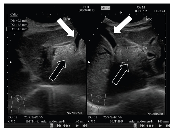
Main gallbladder (white arrow) and cystic structure with multiple stones and debris (black arrow) were confirmed by ultrasonography.
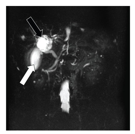
Normal gallbladder (white arrow) and cystic structure (black arrow) were confirmed also by MRCP, but the communication with the biliary tree could not be confirmed.
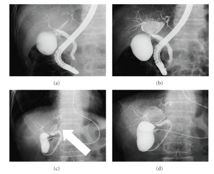
By ERCP the right and left hepatic ducts were displaced (a). With the supplementary injection of radiopaque material, accessory gallbladder could be visualized (b). Cholangiography through ENBD tube conducted the following day showed cystic duct (white arrow) arising from the left hepatic duct (c). Thereafter, an accessory gallbladder was visualized (d).
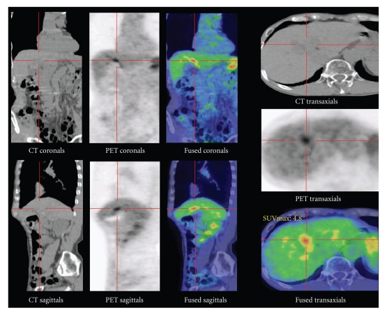
By PET-CT abnormal accumulation of F18-FDP at location considered to be accessory gallbladder could be visualized.
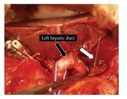
After removing the accessory gallbladder as well as possible, when physiological saline solution was injected into the left hepatic duct, there was a vigorous emission of this solution from the accessory gallbladder (white arrow). After confirming the exit of the cystic duct toward the accessory gallbladder, the exit was ligated.
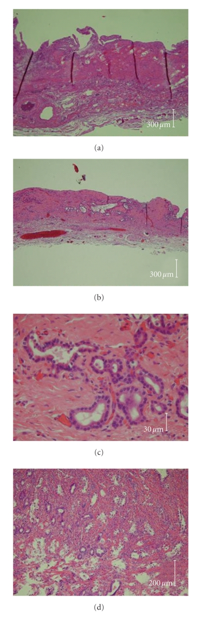
Microscopic image of main gallbladder (a), accessory gallbladders ((b), (c)), and tubular adenocarcinoma in the accessory gallbladder (d). Lobular hyperplasia or adenosis was observed in the accessory gallbladder, but the basic structure of gallbladder (mucosal layer, muscular layer, and submucosal layer) could be seen in both gallbladders. Tubular adenocarcinoma infiltrating into the submucosal layer could be confirmed.
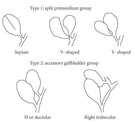
Double gallbladder as classified by Harlaftis et al. [2].
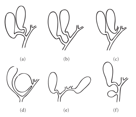
Double gallbladder as classified by Gross [3].
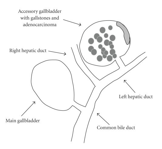
Illustration of our case shows double gallbladder with adenocarcinoma and gallstones arising from the left hepatic duct.
References
-
- Boyden EA. The accessory gall-bladder: an embryological and comparative study of aberrant biliary vesicles occurring in man and the domestic mammals. American Journal of Anatomy. 1926;38(2):177–231.
-
- Harlaftis N, Gray SW, Skandalakis JE. Multiple gallbladders. Surgery Gynecology and Obstetrics. 1977;145(6):928–934. - PubMed
-
- Gross RE. Congenital anomalies of the gallbladder. A review of 148 cases, with report of a double gallbladder. Archives of Surgery. 1969;32:131–162.
-
- Singh B, Ramsaroop L, Allopi L, Moodley J, Satyapal KS. Duplicate gallbladder: an unusual case report. Surgical and Radiologic Anatomy. 2006;28(6):654–657. - PubMed
Publication types
LinkOut - more resources
Full Text Sources
