

Anatomic guidance for ablation: atrial flutter, fibrillation, and outflow tract ventricular tachycardia
- PMID: 20811537
- PMCID: PMC2922872
Anatomic guidance for ablation: atrial flutter, fibrillation, and outflow tract ventricular tachycardia
Abstract
After initial documentation of excellent efficacy with radiofrequency ablation, this procedure is being performed increasingly in more complex situations and for more difficult arrhythmia. In these circumstances, an accurate knowledge of the anatomic basis for the ablation procedure will help maintain this efficacy and improve safety. In this review, we discuss the relevant anatomy for electrophysiology interventions for typical right atrial flutter, atrial fibrillation, and outflow tract ventricular tachycardia. In the pediatric population, maintaining safety is a greater challenge, and here again, knowing the neighboring and regional anatomy of the arrhythmogenic substrate for these arrhythmias may go a long way in preventing complications.
Keywords: ablation; anatomy; atrial fibrillation; atrial flutter; electrophysiology.
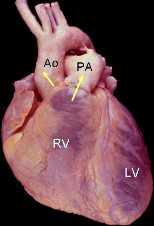
Figures
Similar articles
-
Radiofrequency catheter ablation of atrial arrhythmias. Results and mechanisms.Circulation. 1994 Mar;89(3):1074-89. doi: 10.1161/01.cir.89.3.1074. Circulation. 1994. PMID: 8124793
-
Interventional electrophysiology and its role in the treatment of cardiac arrhythmia.Ann Acad Med Singap. 1998 Mar;27(2):248-54. Ann Acad Med Singap. 1998. PMID: 9663319 Review.
-
Curing reentrant atrial arrhythmias. Targeting protected zones of slow conduction by catheter ablation.J Electrocardiol. 1993;26 Suppl:194-203. J Electrocardiol. 1993. PMID: 8189124
-
[Radiofrequency catheter ablation of atrial flutter and atrial fibrillation].Herz. 1998 Jun;23(4):209-18. doi: 10.1007/BF03044317. Herz. 1998. PMID: 9690109 Review. German.
-
[Catheter ablation of atrial flutter and fibrillation].Rev Esp Cardiol. 1996;49 Suppl 2:55-63. Rev Esp Cardiol. 1996. PMID: 8755697 Review. Spanish.
Cited by
-
Comparison of in-hospital outcomes of patients undergoing catheter ablation for typical versus atypical atrial flutter.J Interv Card Electrophysiol. 2022 Mar;63(2):295-302. doi: 10.1007/s10840-021-00982-4. Epub 2021 Mar 26. J Interv Card Electrophysiol. 2022. PMID: 33770337
-
Outflow tract ventricular tachycardia.Tex Heart Inst J. 2012;39(4):526-8. Tex Heart Inst J. 2012. PMID: 22949769 Free PMC article. Review. No abstract available.
References
-
- Feld GK, et al. Radiofrequency catheter ablation for the treatment of human type 1 atrial flutter. Identification of a critical zone in the reentrant circuit by endocardial mapping techniques. Circulation. 1992;86:1233. - PubMed
-
- Haissaguerre M, et al. Spontaneous initiation of atrial fibrillation by ectopic beats originating in the pulmonary veins. The New England Journal of Medicine. 1998;339:659. - PubMed
-
- Haissaguerre M, et al. Role of catheter ablation for supraventricular tachyarrhythmias, with emphasis on atrial flutter and atrial tachycardia. Current Opinion in Cardiology. 1994;9:40. - PubMed
-
- Pappone C. Atrial fibrillation - a curable condition? European Heart Journal. 2002;23:514. - PubMed
-
- Saad EB, et al. Ablation of atrial fibrillation. Current Cardiology Reports. 2002;4:379. - PubMed
LinkOut - more resources
Full Text Sources
Medical