. 2010 Aug 12;6(8):e1001042.
doi: 10.1371/journal.ppat.1001042.

Early severe inflammatory responses to uropathogenic E. coli predispose to chronic and recurrent urinary tract infection

Affiliations

Early severe inflammatory responses to uropathogenic E. coli predispose to chronic and recurrent urinary tract infection

Thomas J Hannan et al. PLoS Pathog. .

Abstract

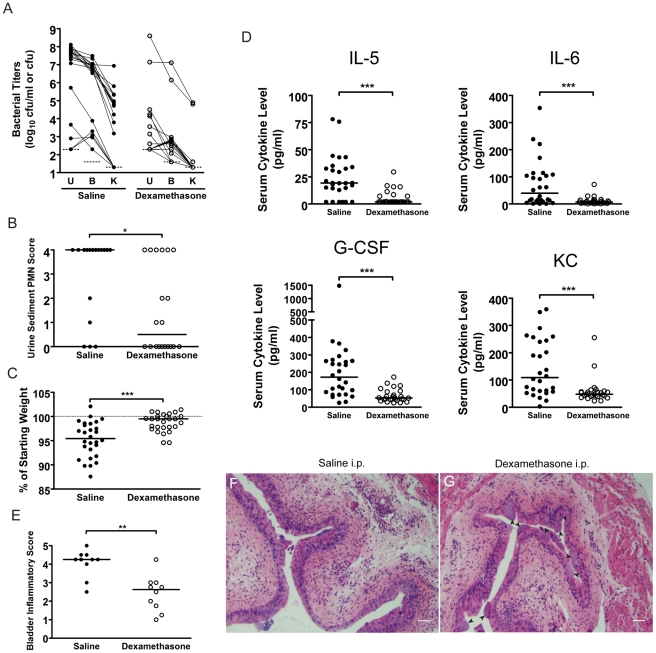
Chronic infections are an increasing problem due to the aging population and the increase in antibiotic resistant organisms. Therefore, understanding the host-pathogen interactions that result in chronic infection is of great importance. Here, we investigate the molecular basis of chronic bacterial cystitis. We establish that introduction of uropathogenic E. coli (UPEC) into the bladders of C3H mice results in two distinct disease outcomes: resolution of acute infection or development of chronic cystitis lasting months. The incidence of chronic cystitis is both host strain and infectious dose-dependent. Further, development of chronic cystitis is preceded by biomarkers of local and systemic acute inflammation at 24 hours post-infection, including severe pyuria and bladder inflammation with mucosal injury, and a distinct serum cytokine signature consisting of elevated IL-5, IL-6, G-CSF, and the IL-8 analog KC. Mice deficient in TLR4 signaling or lymphocytes lack these innate responses and are resistant, to varying degrees, to developing chronic cystitis. Treatment of C3H mice with the glucocorticoid anti-inflammatory drug dexamethasone prior to UPEC infection also suppresses the development of chronic cystitis. Finally, individuals with a history of chronic cystitis, lasting at least 14 days, are significantly more susceptible to redeveloping severe, chronic cystitis upon bacterial challenge. Thus, we have discovered that the development of chronic cystitis in C3H mice by UPEC is facilitated by severe acute inflammatory responses early in infection, which subsequently are predisposing to recurrent cystitis, an insidious problem in women. Overall, these results have significant implications for our understanding of how early host-pathogen interactions at the mucosal surface determines the fate of disease.


Conflict of interest statement

The authors have declared that no competing interests exist.

Figures

Figure 1
Figure 1. C3H/HeN mice develop chronic cystitis in response to UPEC infection in an infectious dose-dependent manner.
A–B, Representative urine bacterial titer time course over 4 wpi with 107 cfu UTI89 KanR in A, C57BL/6J and B, C3H/HeN mice. Solid lines connect the urine titers over time for each individual mouse. Dashed horizontal line represents the cutoff for significant bacteriuria in free catch urines: 104 cfu/ml. C–D, Urine (U), bladder (B) and kidney (K) titers of C, C57BL/6J and D, C3H/HeN mice at 4 wpi with 107 or 108 cfu UTI89 KanR or UTI89, grouped by outcome: resolved bacteriuria (Resolved) or persistent bacteriuria (PB). Solid lines connect the different urine and tissue titers from the same mouse. Dotted horizontal lines show the limits of detection. E–H, Paraffin-embedded bladder sections 4 wpi with UTI89 KanR were examined by E–F, Hematoxylin & eosin (H&E) staining and light microscopy, or G–H, indirect immunofluorescence (IF) microscopy, staining with antibodies against E. coli (green) and uroplakin III (red); nuclei are counterstained with bis-benzimide (blue). Bladder sections from mice that resolved bacteriuria are displayed in panels E and G; from persistently bacteriuric mice in panels F and H. In panel H, arrows indicate luminal clusters of bacteria associated with immune and exfoliated urothelial cells. I–J, Frozen sections of the bladder of a persistently bacteriuric C3H/HeN mouse at 7.5 months post-infection with UTI89 were examined: I, H&E stained section of the trigone region (area of bladder near urethra where ureters enter), J, IF micrograph of a serial section of I, staining for CD45 (red) and the FimH adhesin of E. coli (green); nuclei are counterstained with bis-benzimide (blue). In panels E–J, bars approximate 50µm, “L” indicates bladder lumen, and dashed line denotes the approximate location of the urothelial basement membrane.
Figure 2
Figure 2. The outcome of UPEC infection of the urinary bladder differs between C3H/HeOuJ and C3H/HeJ mice.
C3H/HeOuJ (closed circles) or C3H/HeJ (open circles) mice were infected with 107 cfu UTI89 KanR and followed for 4 wpi. Data were compiled from two independent experiments. A, Urine bacterial titers were assayed over 4 wpi. Solid lines connect the urine titers over time for each individual mouse. Dashed horizontal line represents the cutoff for significant bacteriuria in free catch urines: 104 cfu/ml. B–C, B, Bladder and C, Kidney titers were assayed at 4 wpi; D, Bladders with high bacterial burdens (>104 cfu from panel B) were weighed at 4 wpi. All statistics in panels B, C, and D are the results of a two-tailed Mann-Whitney U test: **, P<0.01, ***, P<0.001 and ns, not significant; horizontal bars indicate median values.
Figure 3
Figure 3. Chronic bladder infection in C3H/HeJ mice is histologically distinct from chronic cystitis in C3H/HeOuJ mice.
Representative paraffin-embedded, fixed bladder sections from C3H/HeOuJ mice with chronic cystitis (22 out of 24 mice; panels A, D, & G), C3H/HeJ mice with high bacterial burdens and evidence of bladder inflammation (5 out of 20 mice; panels B, E, & H), and the remaining C3H/HeJ mice that uniformly lacked evidence of bladder inflammation (15 out of 20 mice; panels C, F, & I) were analyzed at 4 wpi; Bars approximate 50 µm in length. A–F, Sections were stained with hematoxylin & eosin and examined by light microscopy; boxes in panels A–C demarcate boundaries of higher magnification images in panels D–F. G–I, Sections were analyzed by indirect immunofluorescence (IF) microscopy, staining for E. coli (green) and uroplakin III (red), with nuclei counterstained with bis-benzimide (blue). “L” denotes bladder lumen; dashed line indicates approximate location of basement membrane; arrowheads point to urothelial surface bacterial colonization in panel E; and the white arrow points to a mature superficial facet cell containing a possible IBC in panel H.
Figure 4
Figure 4. C3H/HeN mice that develop chronic cystitis can be distinguished from their cage mates by several parameters of infection and host response at 24 hpi.
A–C, Parameters of acute infection and inflammation were assayed in mice that either resolved bacteriuria (R, open circles) or developed persistent bacteriuria (PB, open triangles). A, Urine bacterial titers were assayed at 24 hpi with 107 cfu of UTI89 KanR or UTI89; data is compiled from seven independent experiments (n = 92 mice). B, Urine sediments at 24 hpi with either 107 or 108 cfu of UTI89 KanR or UTI89 were analyzed and assigned a semi-quantitative polymorphonuclear cell (PMN) score from 0–4 as explained in the Materials and Methods; data is compiled from 6 independent experiments (n = 123 mice). C, Mice were weighed at every urine collection time point in most studies. Plot depicts the weight loss or gain at 1 and 28 days post-infection (dpi) with 108 cfu UTI89 KanR; data is compiled from two independent experiments (n = 42 mice). D, Mouse sera collected at 24 hpi with either PBS (Mock) or 108 cfu UTI89 KanR infected mice were analyzed for the presence of 23 cytokines by cytometric bead array as described in the Materials and Methods. UPEC infected mice were then grouped by disease outcome: resolved bacteriuria (Resolved), or persistently bacteriuric (PB). These four cytokines were significantly elevated in PB mice in each of three independent experiments; data shown is from one representative experiment. All statistics are by Mann-Whitney U two-tailed test: *, P<0.05, **, P<0.01, ***, P<0.001 and ns, not significant; horizontal bars indicate median values.
Figure 5
Figure 5. C3H/HeN mice that develop chronic cystitis have severe bladder inflammation at 24 hpi.
Individual C3H/HeN mice were infected with either 107 or 108 cfu of UTI89 and bladders and sera collected at 24 hpi in two independent experiments. Data shown are from one representative experiment. A, Paraffin-embedded, fixed bladder sections were stained with hematoxylin & eosin and examined by light microscopy. Sections were scored from 0–5 to quantify the severity of inflammatory lesions as explained in the Materials and Methods; horizontal bars indicate median values. B, Examples are shown of the disparate response to UPEC infection in two individual mice from the same cage, infected on the same day with the same inoculum (107 cfu UTI89). C–D, Higher magnification images of the two bladders shown in panel B. Inflammatory scores were 3 and 5 for panels C and D, respectively. Bars approximate 50 µm in length. E, Scatter plot analysis of serum IL-5, IL-6, G-CSF, and KC values, as labeled, versus the inflammatory scores of the bladders from the same individual mice. Significance (P) and strength (ρ) of correlations shown were determined by Spearman's rank correlation test.
Figure 6
Figure 6. C3H background severe combined immunodeficient mice have muted acute inflammatory responses to UPEC infection and are resistant to chronic cystitis.
C3H/HeSnJ (closed circles) or C3SnSmn.CB17-Prkcscid/J (C3Hscid, open circles) mice were infected with 108 cfu of UTI89 KanR or UTI89. A, Urine and tissue titers were assayed at 4 wpi; solid lines connect the different urine and tissue titers from the same mouse. B, PMN scores of the urine sediments were determined at 24 hpi. The data shown in panels A and B were compiled from four independent experiments. C, Sera were collected for cytokine analysis at 24 hpi. Data are representative of two independent experiments. All statistics in panels B–C are the results of the Mann-Whitney U test: **, P<0.01, ***, P<0.001 and ns, not significant, horizontal bars indicate median values. D–E, Representative paraffin-embedded, fixed bladder sections from D, C3H/HeSnJ, and E, C3Hscid mice, at 24 hpi were stained with hematoxylin & eosin and examined by light microscopy. Asterisks indicate areas of submucosal edema, arrows indicate apparent IBCs, and bars approximate 50 µm in length.
Figure 7
Figure 7. C3H mice treated with the glucocorticoid anti-inflammatory drug, dexamethasone, just prior to infection have muted acute inflammatory responses to UPEC infection and are protected against chronic cystitis.
C3H/HeN mice were treated with either saline (closed circles) or 200 µg dexamethasone sodium phosphate (open circles) two hours prior to infection with 108 cfu UTI89 KanR. A, urine (U), bladder (B) and kidney (K) titers were determined at 4 wpi; solid lines connect the different urine and tissue titers from the same mouse. B, PMN scores of the urine sediments were determined 24 hpi. A–B, Data are compiled from three independent experiments. C, Percent weight change at 24 hpi was determined. D, Serum cytokine analysis of mice at 24 hpi with 108 cfu of UTI89 KanR is depicted. C–D, Data are compiled from four independent experiments. E–G, C3H/HeN mice pre-treated with either saline or dexamethasone were sacrificed at 24 hpi and paraffin-embedded, fixed bladder sections were stained with hematoxylin & eosin and examined by light microscopy. Semi-quantative bladder inflammatory scores are plotted in panel E, and representative images of bladders from mice treated with F, saline, and G, dexamethasone, are depicted; arrowheads indicate apparent IBCs; bars approximate 50 µm in length. All statistics in panels B–E are the results of the Mann-Whitney U test: *, P<0.05, **, P<0.01, and ***, P<0.001; horizontal bars indicate median values.
Figure 8
Figure 8. C3H/HeN mice with a history of chronic cystitis are more susceptible to chronic cystitis upon challenge infection.
A–D, Mice were infected with 107 UTI89 KanR or mock-infected with PBS and followed for 4 wpi by longitudinal urinalysis in two independent experiments. All mice were treated with antibiotics at 4 wpi and sacrificed 4 weeks after initiation of antibiotic therapy. Paraffin-embedded, fixed bladder sections were stained with hematoxylin & eosin and examined by light microscopy. Representative images of mice that A, were mock-infected, B, had resolved bacteriuria, or C, had persistent bacteriuria are shown; bars approximate 50 µm in length. Statistics are the results of the Mann-Whitney U test: **, P<0.01, and ***, P<0.001. D, Bladders were weighed at sacrifice; horizontal bars indicate median values. E, Experimental time line for panel F: mice were followed by longitudinal urinalysis (each urine collection is indicated by small arrow) for the entire experiment to identify persistently bacteriuric mice prior to antibiotic therapy and after re-infection, and to confirm clearance of bacteriuria with antibiotic therapy. F–H, Combined time courses from two independent experiments each are displayed, charting the UTI89 SpcR urine bacterial titers during the 4 weeks after challenge infection, when antibiotic therapy was initiated at F, 28 days, G, 14 days, or G, 1 day after the initial infection. In all cases, mice were challenged 4 weeks after the initiation of antibiotic therapy. For panels F and G, mice are grouped by the outcome of urinalysis during the initial infection: Mock, mock-infected with PBS at week 0; Resolved Bacteriuria, infected with UTI89 KanR at week 0 and subsequently had at least one time point with <104 cfu/ml UTI89 KanR; or Persistent Bacteriuria, infected with UTI89 KanR at week 0 and had >104 cfu/ml UTI89 KanR for each time point. For panel H, mice are grouped by serum KC value at 24 hpi with UPEC: Mock, mock-infected with PBS at week 0; Resolved Bacteriuria, infected with UTI89 KanR at week 0 and have serum KC in the lower 50% of values at 24 hpi; or Persistent Bacteriuria, infected with UTI89 KanR at week 0 and had serum KC in the upper 50% of values at 24 hpi. Solid lines connect the urine titers over time for each individual mouse. Horizontal dashed lines represent the cutoff for significant bacteriuria in free catch urines: 104 cfu/ml.

References

    1. Arias Ca, Murray Be. Antibiotic-Resistant Bugs In The 21st Century–A Clinical Super-Challenge. N Engl J Med. 2009;360:439–443. - PubMed
    1. Kim Ey, Battaile Jt, Patel Ac, You Y, Agapov E, et al. Persistent Activation Of An Innate Immune Response Translates Respiratory Viral Infection Into Chronic Lung Disease. Nat Med. 2008;14:633–640. - PMC - PubMed
    1. Cassell Gh. Infectious Causes Of Chronic Inflammatory Diseases And Cancer. Emerg Infect Dis. 1998;4:475–487. - PMC - PubMed
    1. Monack Dm, Mueller A, Falkow S. Persistent Bacterial Infections: The Interface Of The Pathogen And The Host Immune System. Nat Rev Microbiol. 2004;2:747–765. - PubMed
    1. Nickel Jc. Management Of Urinary Tract Infections: Historical Perspective And Current Strategies: Part 1–Before Antibiotics. J Urol. 2005;173:21–26. - PubMed

Publication types

MeSH terms