

Lying about the valence of affective pictures: an fMRI study
- PMID: 20811624
- PMCID: PMC2928271
- DOI: 10.1371/journal.pone.0012291
Lying about the valence of affective pictures: an fMRI study
Abstract
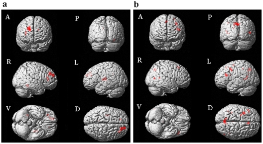
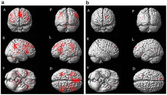
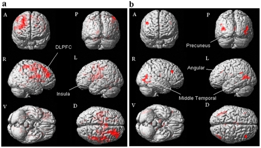
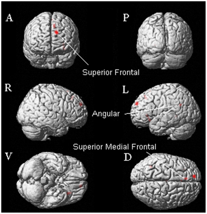
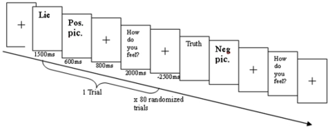
The neural correlates of lying about affective information were studied using a functional magnetic resonance imaging (fMRI) methodology. Specifically, 13 healthy right-handed Chinese men were instructed to lie about the valence, positive or negative, of pictures selected from the International Affective Picture System (IAPS) while their brain activity was scanned by a 3T Philip Achieva scanner. The key finding is that the neural activity associated with deception is valence-related. Comparing to telling the truth, deception about the valence of the affectively positive pictures was associated with activity in the inferior frontal, cingulate, inferior parietal, precuneus, and middle temporal regions. Lying about the valence of the affectively negative pictures, on the other hand, was associated with activity in the orbital and medial frontal regions. While a clear valence-related effect on deception was observed, common neural regions were also recruited for the process of deception about the valence of the affective pictures. These regions included the lateral prefrontal and inferior parietal regions. Activity in these regions has been widely reported in fMRI studies on deception using affectively-neutral stimuli. The findings of this study reveal the effect of valence on the neural activity associated with deception. Furthermore, the data also help to illustrate the complexity of the neural mechanisms underlying deception.
Conflict of interest statement
Figures
Similar articles
-
Let the man choose what to do: Neural correlates of spontaneous lying and truth-telling.Brain Cogn. 2016 Feb;102:13-25. doi: 10.1016/j.bandc.2015.11.007. Epub 2015 Dec 10. Brain Cogn. 2016. PMID: 26685089
-
Neural correlates of deception: lying about past events and personal beliefs.Soc Cogn Affect Neurosci. 2017 Jan 1;12(1):116-127. doi: 10.1093/scan/nsw151. Soc Cogn Affect Neurosci. 2017. PMID: 27798254 Free PMC article.
-
A functional MRI study of deception among offenders with antisocial personality disorders.Neuroscience. 2013 Aug 6;244:90-8. doi: 10.1016/j.neuroscience.2013.03.055. Epub 2013 Apr 9. Neuroscience. 2013. PMID: 23578713
-
What Deception Tasks Used in the Lab Really Do: Systematic Review and Meta-analysis of Ecological Validity of fMRI Deception Tasks.Neuroscience. 2021 Aug 1;468:88-109. doi: 10.1016/j.neuroscience.2021.06.005. Epub 2021 Jun 8. Neuroscience. 2021. PMID: 34111448
-
A Case for Neutrality: Why Neutral Affect is Critical for Advancing Affective Science.Affect Sci. 2023 Aug 21;4(3):458-462. doi: 10.1007/s42761-023-00214-0. eCollection 2023 Sep. Affect Sci. 2023. PMID: 37744984 Free PMC article. Review.
Cited by
-
Neurocognitive mechanisms underlying deceptive hazard evaluation: An event-related potentials investigation.PLoS One. 2017 Aug 9;12(8):e0182892. doi: 10.1371/journal.pone.0182892. eCollection 2017. PLoS One. 2017. PMID: 28793344 Free PMC article.
-
Neural correlates of spontaneous deception: A functional near-infrared spectroscopy (fNIRS)study.Neuropsychologia. 2013 Mar;51(4):704-12. doi: 10.1016/j.neuropsychologia.2012.12.018. Epub 2013 Jan 20. Neuropsychologia. 2013. PMID: 23340482 Free PMC article.
-
Neural mechanisms of deception in a social context: an fMRI replication study.Sci Rep. 2020 Jul 1;10(1):10713. doi: 10.1038/s41598-020-67721-z. Sci Rep. 2020. PMID: 32612101 Free PMC article.
-
Neural correlates of anxiety under interrogation in guilt or innocence contexts.PLoS One. 2020 Apr 9;15(4):e0230837. doi: 10.1371/journal.pone.0230837. eCollection 2020. PLoS One. 2020. PMID: 32271789 Free PMC article.
-
Posterior Midline Activation during Symptom Provocation in Acute Stress Disorder: An fMRI Study.Front Psychiatry. 2014 May 8;5:49. doi: 10.3389/fpsyt.2014.00049. eCollection 2014. Front Psychiatry. 2014. PMID: 24847285 Free PMC article.
References
-
- Ekman P. New York, NY: W W Norton & Co; 2001. Telling lies: Clues to deceit in the marketplace, politics, and marriage.390
-
- Augustine S. Paris: de Brouwer; 1948. “De mendacio.” Opuscules, Vol II, Problémes moraux. pp. 244–245.
-
- Langleben DD, Schroeder L, Maldjian JA, Gur RC, McDonald S, et al. Brain activity during simulated deception: an event-related functional magnetic resonance study. Neuroimage. 2002;15:727–732. - PubMed
-
- Spence SA, Farrow TF, Herford AE, Wilkinson ID, Zheng Y, et al. Behavioural and functional anatomical correlates of deception in humans. Neuroreport. 2001;12:2849–2853. - PubMed
Publication types
MeSH terms
LinkOut - more resources
Full Text Sources
Medical
