. 2010 Nov-Dec;16(11-12):479-90.
doi: 10.2119/molmed.2010.00126. Epub 2010 Aug 27.

Diet restriction inhibits apoptosis and HMGB1 oxidation and promotes inflammatory cell recruitment during acetaminophen hepatotoxicity

Affiliations

Diet restriction inhibits apoptosis and HMGB1 oxidation and promotes inflammatory cell recruitment during acetaminophen hepatotoxicity

Daniel James Antoine et al. Mol Med. 2010 Nov-Dec.

Retraction in

Expression of concern in

Abstract

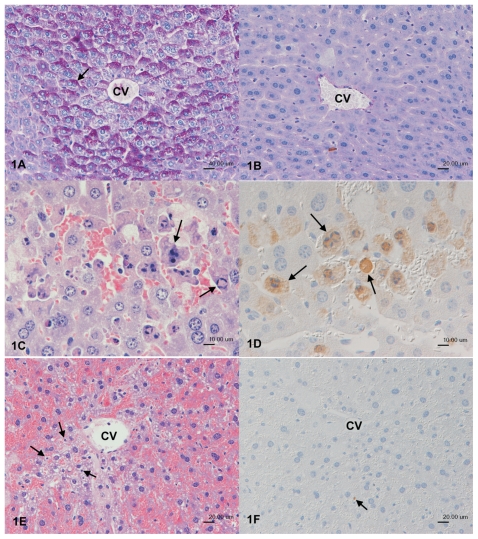
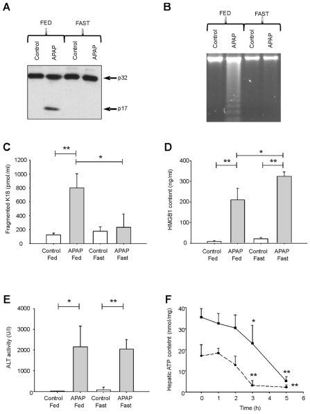
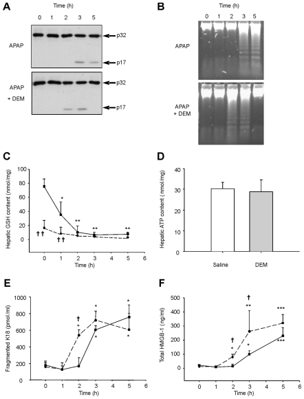
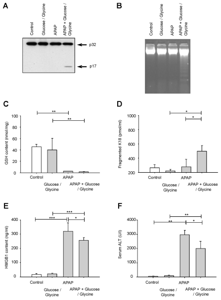
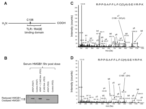
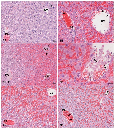
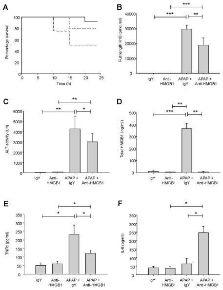
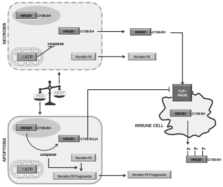
Acetaminophen (APAP) overdose is a major cause of acute liver failure and serves as a paradigm to elucidate mechanisms, predisposing factors and therapeutic interventions. The roles of apoptosis and inflammation during APAP hepatotoxicity remain controversial. We investigated whether fasting of mice for 24 h can inhibit APAP-induced caspase activation and apoptosis through the depletion of basal ATP. We also investigated in fasted mice the critical role played by inhibition of caspase-dependent cysteine 106 oxidation within high mobility group box-1 protein (HMGB1) released by ATP depletion in dying cells as a mechanism of immune activation. In fed mice treated with APAP, necrosis was the dominant form of hepatocyte death. However, apoptosis was also observed, indicated by K18 cleavage, DNA laddering and procaspase-3 processing. In fasted mice treated with APAP, only necrosis was observed. Inflammatory cell recruitment as a consequence of hepatocyte death was observed only in fasted mice treated with APAP or fed mice cotreated with a caspase inhibitor. Hepatic inflammation was also associated with loss in detection of serum oxidized-HMGB1. A significant role of HMGB1 in the induction of inflammation was confirmed with an HMGB1-neutralizing antibody. The differential response between fasted and fed mice was a consequence of a significant reduction in basal hepatic ATP, which prevented caspase processing, rather than glutathione depletion or altered APAP metabolism. Thus, the inhibition of caspase-driven apoptosis and HMGB1 oxidation by ATP depletion from fasting promotes an inflammatory response during drug-induced hepatotoxicity/liver pathology.


Figures

Figure 1
Figure 1
Histological determination of the effect of fasting on APAP-induced hepatocyte apoptosis in mice. (A, B) Control mice, PAS reaction. (A) In control mice with free access to food, the vast majority of centrilobular hepatocytes exhibit cytoplasmic glycogen (arrow). (B) Mice fasted for 24 h do not exhibit glycogen within hepatocytes. Bars = 20 μm. (C–F) 3 hours after APAP treatment (530 mg/kg). In animals with free access to food (C, D), centrilobular cell loss was observed (average score 1.1). There were apoptotic cells that could be identified based on their morphology in a hematoxylin and eosin–stained section (arrows, C) and the expression of cleaved caspase-3 (arrows, D). Bars = 10 μm. Animals that had been fasted for 24 h before APAP administration showed more extensive hepatocyte loss (average score 1.6). Necrotic hepatocytes were identified in the hematoxylin and eosin–stained section (arrows, E). There was no evidence of cleaved caspase-3 expression by hepatocytes, and the only positive apoptotic cells were scattered leukocytes or Kupffer cells (arrow, F). Bars = 20 μm. Figures are representative of six animals per group carried out in three independent investigations. CV, central vein.
Figure 2
Figure 2
The effect of 24-h fasting of mice on the (A) processing of hepatic procaspase-3 (p32) to the active form (p17) by Western blot and (B) hepatic DNA laddering induced by APAP (530 mg/kg; 5 h). The effect of 24 h-fasting of mice on the serum level of (C) K18 apoptotic fragments (pmol/mL), (D) total HMGB1 content (ng/mL) and (E) ALT activity (U/L) after APAP treatment (530 mg/kg; 5 h). (F) The effect of APAP (530 mg/kg) on hepatic ATP content was determined over 0–5 h in fed (solid line) and fasted mice (dotted line). Figures are representative and data is given as mean ± SD of six mice per group carried out in three independent investigations. Statistical significance was assigned as *P < 0.05, **P < 0.01 relative to time-matched controls.
Figure 3
Figure 3
The effect of DEM predepletion (dotted line) of hepatic GSH on APAP-induced (530 mg/kg; 0–5 h) apoptotic and necrotic cell death compared with saline treatment (solid line) in fed male CD-1 mice. Representative (A) Western analysis of hepatic processing of procaspase-3 (p32) to the active form (p17) and (B) agarose gel analysis of hepatic DNA laddering. The effect of GSH predepletion on APAP-induced (C) hepatic GSH depletion, (D) hepatic ATP depletion and serum levels of (E) caspase-cleaved K18 fragments (pmol/mL) and (F) serum HMGB1 content (ng/mL) was determined simultaneously 0–5 h after APAP treatment. Data are given as mean ± SD of six mice per group carried out in three independent investigations. Statistical significance was assigned relative to vehicle treated controls (*P < 0.05, **P < 0.01 and ***P < 0.005) or between DEM and saline pretreated mice (†P < 0.05) at that time point.
Figure 4
Figure 4
The effect of glucose and glycine administration (1000/100 mg/kg; 1 h pre-APAP) to fasted male CD-1 mice on APAP-induced (530 mg/kg; 5 h) apoptosis and necrosis. (A) Processing of hepatic procaspase-3, (B) hepatic DNA laddering, (C) hepatic GSH depletion (nmol/mg), (D) serum caspase-mediated K18 fragment level (pmol/mL), (E) total serum HMGB1 content (ng/mL) and (F) serum ALT activity (U/L). Figures are representative and data is given as mean ± SD of six mice per group carried out in three independent investigations. Statistical significance was assigned relative to vehicle-treated controls or between APAP alone and APAP + glucose/glycine (*P < 0.05, **P < 0.01 and ***P < 0.005).
Figure 5
Figure 5
Characterization of the cysteine 106 HMGB1 oxidation status with the sera of APAP (530 mg/kg; 5 h) dosed mice. (A) Schematic overview of the location of cysteine 106 within the cytokine domain of murine HMGB1, which is characterized by MS/MS. (B) Nonreducing SDS-PAGE separation and Western analysis of reduced and oxidized HMGB1 isolated by immunoprecipitation from the sera of fed and fasted APAP-dosed mice (530 mg/kg; 5 h) and characterized by LC-MS/MS. HMGB1 was also isolated from Z-VAD.fmk pretreated APAP-dosed mice. (C) MS/MS analysis of oxidized murine HMGB1 peptide 97–112 from a fed APAP-treated mouse containing the cysteine 106 sulfonic acid. (D) MS/MS analysis of reduced murine HMGB1 peptide 97–112 containing the cysteine 106 thiol from an APAP-treated mouse that was prefasted for 24 h. MS/MS and Western figures are representative of six mice per group carried out in three independent investigations.
Figure 6
Figure 6
Histological determination of the effect of fasting and Z-VAD.fmk and anti-HMGB1 coapplication on APAP-induced hepatic changes in mice, 24 h after treatment. (A) In fed mice, there is no evidence of hepatocyte necrosis and the liver looks unaltered apart from an increased number of mitotic hepatocytes (arrow). There is no evidence of inflammatory cell recruitment into the liver. (B, C) Mice fasted for 24 h before treatment (B) and mice treated with APAP + Z-VAD.fmk show evidence of marked hepatocyte loss (score 3) and exhibit neutrophils in the lumen and rolling along endothelial cells (arrows) of central and portal veins, indicating leukocyte recruitment into the liver. (D) Mouse fasted for 24 h before APAP treatment. Closer view of a central vein with numerous neutrophils rolling along endothelial cells (arrows). (E). Treatment of fasted mice with APAP and anti-HMGB1 is associated with evidence of marked hepatocyte loss (score 3), but not of inflammatory cell recruitment into the liver. (F) After treatment of fasted mice with APAP and IgY; however, evidence of marked hepatocyte loss (score 3) and evidence of leukocyte recruitment, represented by neutrophils rolling along endothelial cells (arrows) of central and portal veins, is seen. Hematoxylin and eosin stain. Bars = 20 μm. PA, portal area; CV, central vein. Figures are representative of six animals per group carried out in three independent investigations.
Figure 7
Figure 7
Effect of HMGB1 neutralization on APAP (530 mg/kg; 24 h) hepatotoxicity. Mice were treated with an HMGB1-neutralizing antibody or a control IgY antibody 2 h after APAP treatment and (A) percent (%) survival over 24 h (Fed mice + APAP, solid line; fasted mice + APAP/IgY, large dashed line; fasted mice + APAP/anti-HMGB1, small dashed line), (B) serum full-length K18 level (pmol/mL), (C) serum ALT activity (U/L), (D) serum total HMGB1 content, (E) serum TNF-α level (pg/mL) and (F) serum IL-6 level (pg/mL) were recorded simultaneously within the same group of mice. Data are representative of mean ± SD of six mice per group carried out in three independent investigations. Statistical significance was assigned between groups as appropriate. *P < 0.05, **P < 0.01 and ***P < 0.005.
Figure 8
Figure 8
Schematic overview summarizing the findings within this investigation. APAP-induced hepatic apoptosis and necrosis in the fed CD-1 mouse model, which can be quantified by the serum biomarker K18. This is accompanied by the caspase-dependent oxidation of HMGB1 (HMGB1-SO3H), which prevents an inflammatory response. The fasting of mice before APAP induces a depletion in basal hepatic ATP levels, which does not permit apoptosis activation and HMGB1 oxidation. The resulting reduced HMGB1 (HMGB1-SH) activates inflammatory cells and promotes the hepatic inflammatory response after hepatocyte death, leading to exacerbated injury.

Similar articles

Cited by

References

    1. Gunawan BK, Kaplowitz N. Mechanisms of drug-induced liver disease. Clin Liver Dis. 2007;11:459–75. - PubMed
    1. Hawton K, et al. UK legislation on analgesic packs: before and after study of long term effect on poisonings [erratum appears in BMJ. (2004) 329:1159] BMJ. 2004;329:1076. - PMC - PubMed
    1. Dahlin DC, Miwa GT, Lu AY, Nelson SD. N-acetyl-p-benzoquinone imine: a cytochrome P-450-mediated oxidation product of acetaminophen. Proc Natl Acad Sci U S A. 1984;81:1327–31. - PMC - PubMed
    1. Mitchell JR, Jollow DJ, Potter WZ, Gillette JR, Brodie BB. Acetaminophen-induced hepatic necrosis. IV. Protective role of glutathione. J Pharmacol Exp Ther. 1973;187:211–7. - PubMed
    1. Mitchell JR, et al. Acetaminophen-induced hepatic necrosis, I: role of drug metabolism. J Pharmacol Exp Ther. 1973;187:185–94. - PubMed

Publication types