. 2010 Oct;37(4):983-91.
doi: 10.3892/ijo_00000749.

Biological effects of induced MYCN hyper-expression in MYCN-amplified neuroblastomas

Affiliations

Biological effects of induced MYCN hyper-expression in MYCN-amplified neuroblastomas

Jaime Torres et al. Int J Oncol. 2010 Oct.

Abstract

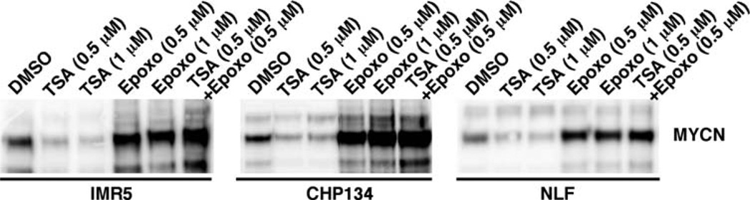
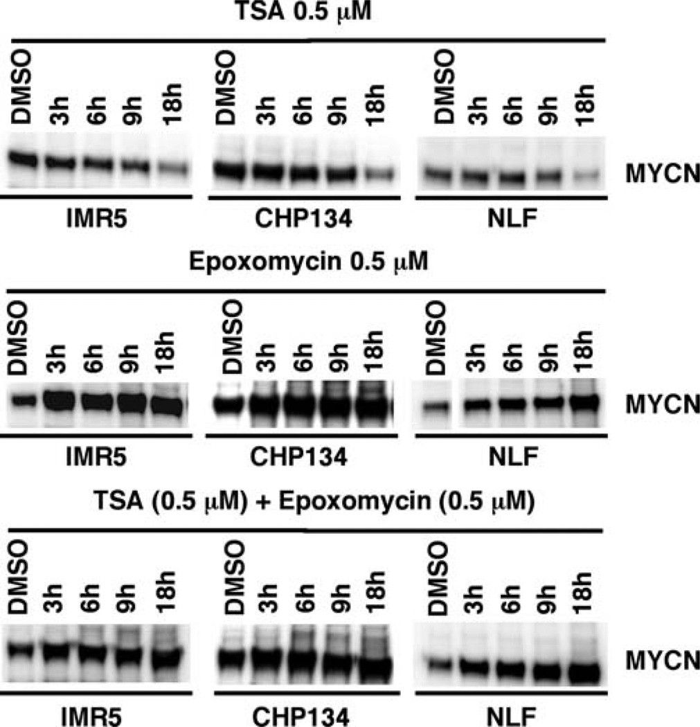
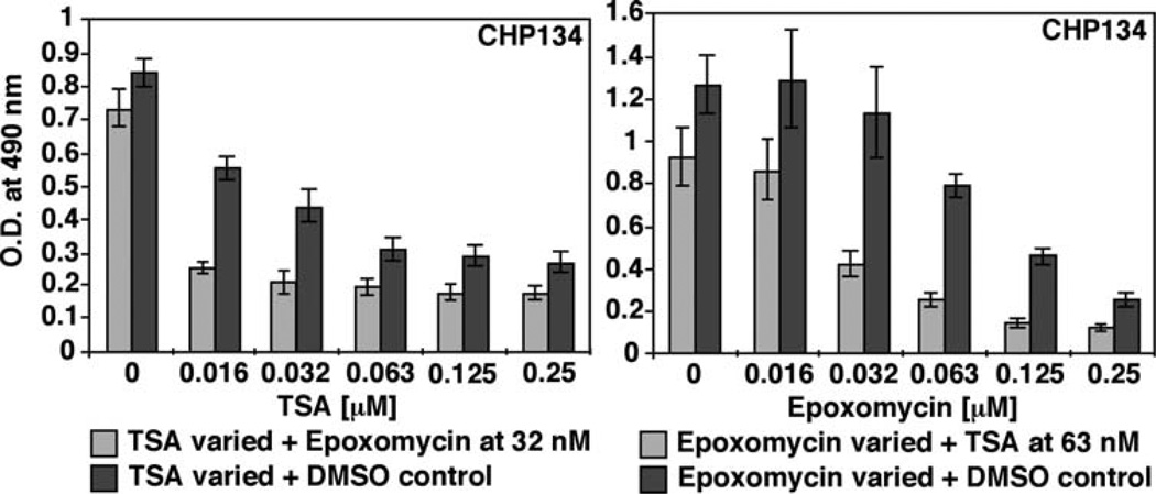
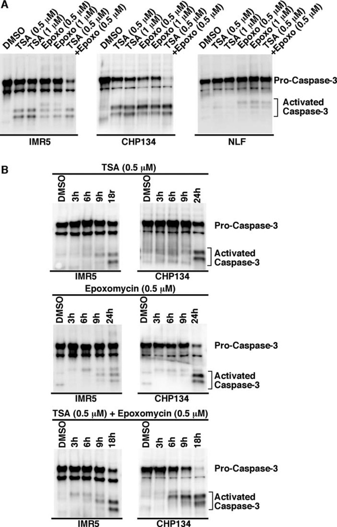
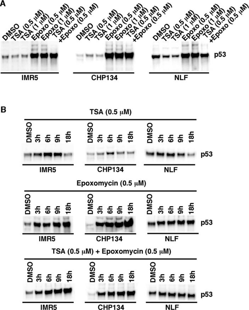
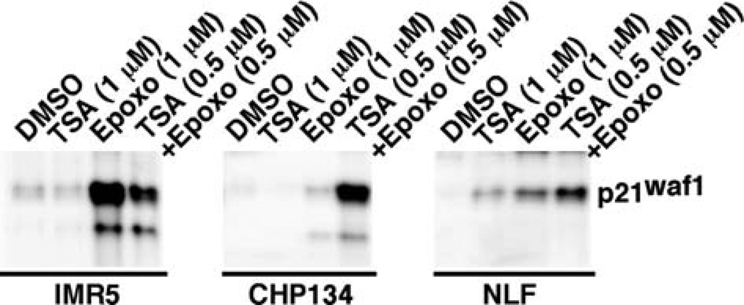
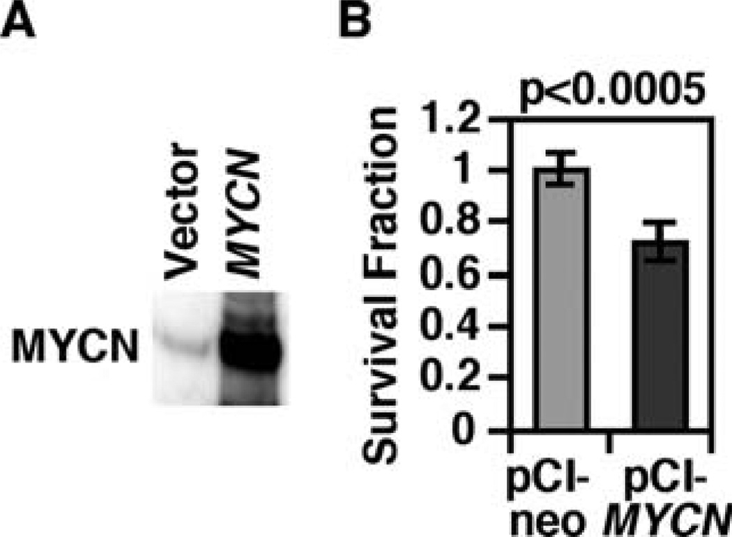
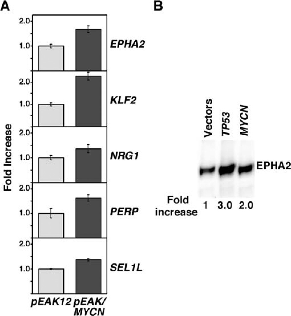
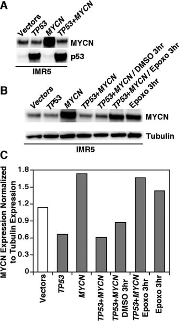
Neuroblastoma is a childhood malignancy of the sympathetic nervous system. The tumor exhibits two different phenotypes: favorable and unfavorable. MYCN amplification is associated with rapid tumor progression and the worst neuroblastoma disease outcome. We have previously reported that inhibitors of histone deacetylase (HDAC) and proteasome enhance favorable neuroblastoma gene expression in neuroblastoma cell lines and inhibit growth of these cells. In this study, we investigated the effect of trichostatin A or TSA (an HDAC inhibitor), and epoxomycin (a proteasome inhibitor) on MYCN and p53 expression in MYCN-amplified neuroblastoma cells. It was found that TSA down-regulated MYCN expression, but Epoxomycin and the TSA/Epoxomycin combination led to MYCN hyper-expression in MYCN-amplified neuroblastoma cell lines. Despite their contrasting effects on MYCN expression, TSA and Epoxomycin caused growth suppression and cell death of the MYCN-amplified cell lines examined. Consistent with these data, forced hyper-expression of MYCN in MYCN-amplified IMR5 cells via transfection resulted in growth suppression and the increased expression of several genes known to suppress growth or induce cell death. Furthermore, Epoxomycin as a single agent and its combination with TSA enhance p53 expression in the MYCN-amplified neuroblastoma cell lines. Unexpectedly, co-transfection of TP53 and MYCN in IMR5 cells resulted in high p53 expression but a reduction of MYCN expression. Together our data suggest that either down regulation or hyper-expression of MYCN results in growth inhibition and/or apoptosis of MYCN-amplified neuroblastoma cells. In addition, elevated p53 expression has a suppressive effect on MYCN expression in these cells.


Figures

Figure 1
Figure 1
TSA and Epoxomycin have contrasting effects on MYCN expression in MYCN-amplified neuroblastoma cell lines (IMR5, CHP134 and NLF). IMR5, CHP134, and NLF were treated with TSA (0.5 or 1 µM), Epoxomycin (0.5 or 1 µM) and the combination TSA (0.5 µM) plus Epoxomycin (0.5 µM) for 18 h. The cells were harvested and subjected to Western blot analysis. Total protein (5 µg) was loaded per lane. The MYCN-specific monoclonal antibody, NCM II 100, was used to detect expression of MYCN (11). The experiments were repeated at least three times to demonstrate the reproducibility of these results.
Figure 2
Figure 2
Time course studies on the effect of TSA, Epoxomycin, and their combination on MYCN expression in MYCN-amplified neuroblastoma cell lines (IMR5, CHP134 and NLF). IMR5, CHP134, and NLF were treated with TSA (0.5 µM), Epoxomycin (0.5 µM) and the combination of TSA (0.5 µM) plus Epoxomycin (0.5 µM) for 3, 6, 9 and 18 h. The cells were harvested and subjected to Western blot analysis. The experiments were done as described in Fig. 1.
Figure 3
Figure 3
TSA and Epoxomycin as single agents or in combination significantly suppress growth of CHP134. CHP134 cells were treated with TSA and Epoxomycin as single agents (dark gray) or in combination (light gray) at the concentrations indicated. After 24 h of the treatments, an MTS assay was done to determine the effect of the experimental drugs on growth of the neuroblastoma cell lines examined. IMR5 and NLF showed similar results (data not shown).
Figure 4
Figure 4
(A) TSA and Epoxomycin as single agents or in combination activate Caspase-3 in IMR5 and CHP134 (retinoid-sensitive lines), whereas Epoxomycin and the combination weakly activate Caspase-3 in NLF. The neuroblastoma cell lines were treated with TSA, Epoxomycin, and the combination for 18 h at the concentrations indicated. Caspase-3 activation was investigated in the drug-treated cells by Western blot analysis using antibodies that could detect both pro and active forms of Caspase-3 (Stressgen, AAP-113). Total protein (10 µg) was loaded per lane. (B) The effect of TSA and/or Epoxomycin on Caspase-3 activation in time course studies. Neuroblastoma cell lines indicated were treated at various time points with TSA, Epoxomycin, and the combination as shown. Caspase-3 activation was investigated in the drug-treated cells as described in (A).
Figure 5
Figure 5
(A) The effect of TSA and Epoxomycin on p53 expression in MYCN-amplified neuroblastoma cell lines (IMR5, CHP134 and NLF). IMR5, CHP134, and NLF were treated with TSA (0.5 or 1 µM), Epoxomycin (0.5 or 1 µM) and the combination of TSA (0.5 µM) plus Epoxomycin (0.5 µM) for 18 h. (B) Time course studies on the effect of the tested drugs on p53 expression. IMR5, CHP134, and NLF were treated with TSA (0.5 µM), Epoxomycin (0.5 µM) and the combination of TSA (0.5 µM) plus Epoxomycin (0.5 µM) for 3, 6, 9 and 18 or 24 h as shown. The cells were harvested and subjected to Western blot analysis. Total protein (10 µg) was loaded per lane. A p53-specific monoclonal antibody, DO-1 (Calbiochem), was used to detect expression of p53. The experiments were repeated at least three times to demonstrate the reproducibility of these results.
Figure 6
Figure 6
The effect of TSA and Epoxomycin on p21waf1 expression in MYCN-amplified neuroblastoma cell lines (IMR5, CHP134 and NLF). IMR5, CHP134, and NLF were treated with TSA (1 µM), Epoxomycin (1 µM) and the combination of TSA (0.5 µM) plus Epoxomycin (0.5 µM) for 18 h. The cells were harvested and subjected to Western blot analysis using an anti-p21waf1 antibody (OP64, Calbiochem). Total protein (20 µg) was loaded per lane. The experiments were repeated at least three times.
Figure 7
Figure 7
(A and B) Forced MYCN expression suppresses growth of IMR5 cells. (A) A full-length cDNA of MYCN was cloned into a eukaryotic expression vector, pCI-neo. IMR5 cells were transfected with either the vector control or the pCI/MYCN cDNA construct by electroporation. Twenty-four h after transfection, the cells were harvested and subjected to Western blot analysis. Five micrograms of total protein were loaded per lane. The MYCN-specific monoclonal antibody, NCM II 100, was used to detect the expression of MYCN. It should be noted that the expression of MYCN in the transfected cells was as high as that shown in the combination TSA plus Epoxomycin treatment (see Fig. 1). (B) To examine the effect of ectopic expression of MYCN in IMR5, the resulting transfectants were selected by neomycin (500 µg/ml) for 5 days, and an MTS assay (a soluble form of MTT assay) was done.
Figure 8
Figure 8
(A) Enhanced expression of genes involved in growth and tumor suppression by MYCN hyper-expression induced by transfection of MYCN in MYCN-amplified IMR5 cells. IMR5 cells were transfected with pEAK12 vector control or pEAK12/MYCN. The cells were harvested two days after transfection. Transfections were performed in duplicate for vector control and triplicate for pEAK12/MYCN. RNAs were prepared from the transfected cells, and pools of RNA from each transfection condition were used in reverse transcription. Expression of genes of interest was examined using TaqMan real-time PCR. Expression levels of EPHA2, KLF2, NRG1, PERP, SEL1L were presented as fold increase in the MYCN transfected IMR5 cells over the vector control. (B) Ectopic expression of either MYCN or p53 induces EPHA2 expression in IMR5 cells. EPHA2 is a known downstream effector of p53 (20). We therefore included TP53-transfected IMR5 cells as a comparative control in this experiment. pCI/MYCN and the TP53 construct, SN3, were transfected to IMR5 by electroporation. Twenty-four h later, the cells were harvested and subjected to Western blot analysis using the mouse monoclonal antibody anti-EPHA2 clone D7 (Millipore). Twenty micrograms of total protein were loaded per lane.
Figure 9
Figure 9
Elevated p53 expression suppresses MYCN expression in a proteasome-dependent fashion. (A) IMR5 cells were transfected with control vectors, the wild-type TP53 construct, the MYCN construct or the combination of TP53 and MYCN. After 24 h of transfection, MYCN and p53 expression was determined by Western blot assay. Total protein (5 µg) was loaded per lane. (B) Lane 1–4: IMR5 cells were treated as described in (A). Lanes 5–6: The double TP53/MYCN transfectants were also treated with either the solvent control DMSO or Epoxomycin (1 µM) for the last 3 h. Lane 7 (the last lane): IMR5 cells treated with only Epoxomycin (1 µM) for 3 h were included as a control. (C) Normalized expression levels of MYCN against tubulin in the conditions shown in (B).

References

    1. Moreau LA, McGrady P, London WB, Shimada H, Cohn SL, Maris JM, Diller L, Look AT, George RE. Does MYCN amplification manifested as homogeneously staining regions at diagnosis predict a worse outcome in children with neuroblastoma? A Children's Oncology Group study. Clin Cancer Res. 2006;12:5693–5697. - PubMed
    1. Seeger RC, Brodeur GM, Sather H, Dalton A, Siegel SE, Wong KY, Hammond D. Association of multiple copies of the N-myc oncogene with rapid progression of neuroblastomas. New Engl J Med. 1985;313:1111–1116. - PubMed
    1. Brodeur GM, Seeger RC, Schwab M, Varmus HE, Bishop JM. Amplification of N-myc in untreated human neuroblastomas correlates with advanced disease stage. Science. 1984;224:1121–1124. - PubMed
    1. Thiele CJ, Reynolds CP, Israel MA. Decreased expression of N-myc precedes retinoic acid-induced morphological differentiation of human neuroblastoma. Nature. 1985;313:404–406. - PubMed
    1. Kang JH, Rychahou PG, Ishola TA, Qiao J, Evers BM, Chung DH. MYCN silencing induces differentiation and apoptosis in human neuroblastoma cells. Biochem Biophys Res Commun. 2006;351:192–197. - PMC - PubMed

Publication types

MeSH terms