The cerebellum and basal ganglia are interconnected
- PMID: 20811947
- PMCID: PMC3325093
- DOI: 10.1007/s11065-010-9143-9
The cerebellum and basal ganglia are interconnected
Abstract
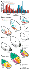
The cerebellum and the basal ganglia are major subcortical nuclei that control multiple aspects of behavior largely through their interactions with the cerebral cortex. Discrete multisynaptic loops connect both the cerebellum and the basal ganglia with multiple areas of the cerebral cortex. Interactions between these loops have traditionally been thought to occur mainly at the level of the cerebral cortex. Here, we review a series of recent anatomical studies in nonhuman primates that challenge this perspective. We show that the anatomical substrate exists for substantial interactions between the cerebellum and the basal ganglia. Furthermore, we discuss how these pathways may provide a useful framework for understanding cerebellar contributions to the manifestation of two prototypical basal ganglia disorders, Parkinson's disease and dystonia.
Conflict of interest statement
Figures


Similar articles
-
The basal ganglia and the cerebellum: nodes in an integrated network.Nat Rev Neurosci. 2018 Jun;19(6):338-350. doi: 10.1038/s41583-018-0002-7. Nat Rev Neurosci. 2018. PMID: 29643480 Free PMC article. Review.
-
The basal ganglia: motor and cognitive relationships in a clinical neurobehavioral context.Rev Neurosci. 2013;24(1):9-25. doi: 10.1515/revneuro-2012-0067. Rev Neurosci. 2013. PMID: 23241666 Review.
-
Proceedings of the workshop on Cerebellum, Basal Ganglia and Cortical Connections Unmasked in Health and Disorder held in Brno, Czech Republic, October 17th, 2013.Cerebellum. 2015 Apr;14(2):142-50. doi: 10.1007/s12311-014-0595-y. Cerebellum. 2015. PMID: 25205331 Free PMC article.
-
A new twist on the anatomy of dystonia: the basal ganglia and the cerebellum?Neurology. 2006 Nov 28;67(10):1740-1. doi: 10.1212/01.wnl.0000246112.19504.61. Neurology. 2006. PMID: 17130402 No abstract available.
-
The cerebellum communicates with the basal ganglia.Nat Neurosci. 2005 Nov;8(11):1491-3. doi: 10.1038/nn1544. Epub 2005 Oct 2. Nat Neurosci. 2005. PMID: 16205719
Cited by
-
Torsin A Localization in the Mouse Cerebellar Synaptic Circuitry.PLoS One. 2013 Jun 19;8(6):e68063. doi: 10.1371/journal.pone.0068063. Print 2013. PLoS One. 2013. PMID: 23840813 Free PMC article.
-
The Neurodevelopmental Hypothesis of Huntington's Disease.J Huntingtons Dis. 2020;9(3):217-229. doi: 10.3233/JHD-200394. J Huntingtons Dis. 2020. PMID: 32925079 Free PMC article. Review.
-
Structural and connectivity parameters reveal spared connectivity in young patients with non-progressive compared to slow-progressive cerebellar ataxia.Front Neurol. 2023 Oct 30;14:1279616. doi: 10.3389/fneur.2023.1279616. eCollection 2023. Front Neurol. 2023. PMID: 37965172 Free PMC article.
-
Cerebral activations related to ballistic, stepwise interrupted and gradually modulated movements in Parkinson patients.PLoS One. 2012;7(7):e41042. doi: 10.1371/journal.pone.0041042. Epub 2012 Jul 23. PLoS One. 2012. PMID: 22911738 Free PMC article.
-
Striatal cholinergic dysfunction as a unifying theme in the pathophysiology of dystonia.Prog Neurobiol. 2015 Apr;127-128:91-107. doi: 10.1016/j.pneurobio.2015.02.002. Epub 2015 Feb 17. Prog Neurobiol. 2015. PMID: 25697043 Free PMC article. Review.
References
-
- Alexander GE, Crutcher MD. Functional architecture of basal ganglia circuits: neural substrates of parallel processing. Trends in Neurosciences. 1990;13(7):266–271. - PubMed
-
- Alexander GE, DeLong MR, Strick PL. Parallel organization of functionally segregated circuits linking the basal ganglia and cortex. Annual Review of Neuroscience. 1986;9:357–381. - PubMed
-
- Allen GI, Tsukahara N. Cerebrocerebellar communication systems. Physiological Review. 1974;54(4):957–1006. - PubMed
-
- Amaral DG, Schumann CM, Nordahl CW. Neuroanatomy of autism. Trends in Neurosciences. 2008;31(3):137–145. - PubMed
Publication types
MeSH terms
Grants and funding
LinkOut - more resources
Full Text Sources

