Review
. 2011 Apr 15;14(8):1551-85.
doi: 10.1089/ars.2010.3228. Epub 2011 Jan 8.

Aging and immune function: molecular mechanisms to interventions

Affiliations
Review

Aging and immune function: molecular mechanisms to interventions

Subramaniam Ponnappan et al. Antioxid Redox Signal. .

Abstract

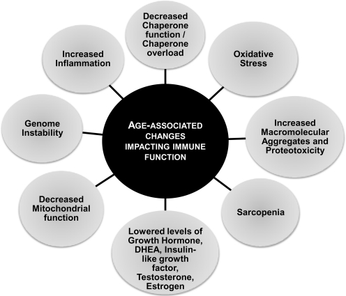
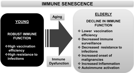
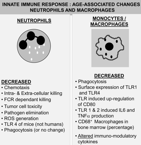
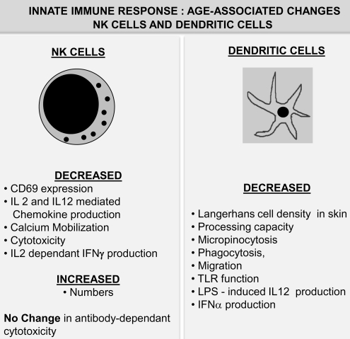
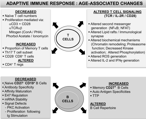
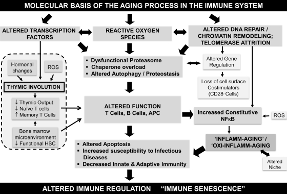
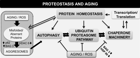
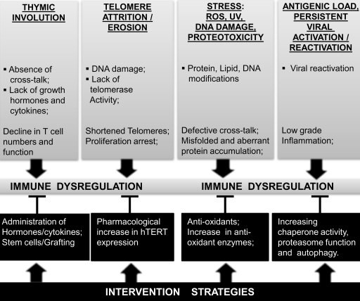
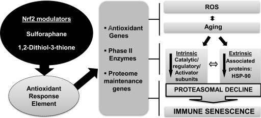
Abstract The immune system of an organism is an essential component of the defense mechanism aimed at combating pathogenic stress. Age-associated immune dysfunction, also dubbed "immune senescence," manifests as increased susceptibility to infections, increased onset and progression of autoimmune diseases, and onset of neoplasia. Over the years, extensive research has generated consensus in terms of the phenotypic and functional defects within the immune system in various organisms, including humans. Indeed, age-associated alterations such as thymic involution, T cell repertoire skewing, decreased ability to activate naïve T cells and to generate robust memory responses, have been shown to have a causative role in immune decline. Further, understanding the molecular mechanisms underlying the generation of proteotoxic stress, DNA damage response, modulation of ubiquitin proteasome pathway, and regulation of transcription factor NFκB activation, in immune decline, have paved the way to delineating signaling pathways that cross-talk and impact immune senescence. Given the role of the immune system in combating infections, its effectiveness with age may well be a marker of health and a predictor of longevity. It is therefore believed that a better understanding of the mechanisms underlying immune senescence will lead to an effective interventional strategy aimed at improving the health span of individuals. Antioxid. Redox Signal. 14, 1551-1585.


Figures

FIG. 1.
FIG. 1.
An overview of alterations impacting immune status in humans with advancing age. Key changes that have been reported to occur in humans during advancing age, affecting the immune status and resulting in overall decline in immune function, are depicted. DHEA, dehydroepiandrosterone.
FIG. 2.
FIG. 2.
A synopsis of immune senescence and its role in host defense. A summary of observed pathological manifestations attributed to altered immune function during advancing age is depicted. Immune dysfunction in the elderly that often results in increased morbidity and mortality due to infections is contrasted with robust immune function in the young that includes an effective response to vaccination and an enhanced ability to resist infections.
FIG. 3.
FIG. 3.
Projected increase (%) in aged population during the period 2008–2040. The proportion of elderly individuals aged 65 years and older is expected to increase during the next three decades. Projected increase in the total world population is contrasted with that of the elderly population (65 years and older or 80 years and older) during the next three decades using data obtained from the U.S. Census Bureau report. During the period from 2008 to 2040, the population of individuals aged 65 years and above is projected to increase by 160%, whereas that of individuals aged 80 years and older will be over 230%.
FIG. 4.
FIG. 4.
Age-associated alterations in innate immunity: I. Neutrophils and macrophages. Neutrophils: A summary of reported changes in neutrophil function resulting in alterations in innate immune responses during advanced age in humans and mice is presented. Age-associated alteration in neutrophils manifests as a decline in most functional responses with a consistent and significant increase in pro-inflammatory cytokine production. Macrophages: A summary of age-associated alterations in macrophages is depicted. While a minimal decline in numbers is reported in some studies, consistent functional deficits ranging from decreased TLR expression to phagocytosis is observed. Additionally, TLR-induced pro-inflammatory cytokine production has been demonstrated to decline with age. FCR, Fc receptor; ROS, reactive oxygen species; TLR, toll-like receptor.
FIG. 5.
FIG. 5.
Age-associated alterations in innate immunity: II. NK cells and dendritic cells. NK cells: Aging is not only accompanied by a significant increase in NK cell numbers, but significant functional deficits have also been reported to occur in these cells. Dendritic cells: A compilation of changes in function of the dendritic cell population during advanced age in humans is presented. Unlike other immune cells, numbers of dendritic cells remain relatively unchanged with age. However, dendritic cell functions, including antigen processing, antigen presentation, and phagocytosis, appear to be significantly affected by advanced age. IL, interleukin; NK, natural killer.
FIG. 6.
FIG. 6.
Summary of age-associated alterations in adaptive immunity: T lymphocytes and B lymphocytes. Observed alterations in B and T lymphocyte phenotypic and functional attributes during advancing age in humans that contribute to the immune senescence phenotype are presented. B lymphocytes: While both signaling within B cells and antibody affinity maturation decline in advanced age, increases in auto-antigen specificities as well as memory B cell numbers appear to accompany aging. T lymphocytes: Defects ranging from T cell emigration to the periphery to aberrant signaling within the TCR activation pathway have been demonstrated to be largely responsible for the reported age-associated functional deficits. IFN, interferon; NFAT, nuclear factor of activated T cells; NFκB, nuclear factor kappa B; TCR, T cell receptor.
FIG. 7.
FIG. 7.
An outline of the biochemical and molecular mechanisms reported to impact immune function during advancing age, resulting in immune senescence. A schematic representation of the molecular basis of aging in the immune system is presented. Reported studies implicate a significant role for altered transcription factor induction, increased generation of ROS, altered DNA repair mechanisms, and telomere attrition in the dysfunctional immune system. Cross-talk and feedback regulation among these mechanisms appear to manifest as immune dysfunction, and impact a series of functional mechanisms and targets within the immune system that may account for increased susceptibility to infections and the development of an inflammatory phenotype. APC, antigen presenting cell.
FIG. 8.
FIG. 8.
Schematic representation of mechanisms reported to modulate proteostasis during aging. Proteostatic mechanisms and the control of protein stability during aging are presented. Factors such as ROS, accumulation of aberrant proteins, failure of the ubiquitin proteasome pathway, chaperone overload, and decline in autophagy have all been reported to contribute to some extent to the decline in protein homeostasis and to overall functional alteration during advanced age. Recent studies highlight the important roles for BAG3, sequestosome, and HDAC6 in regulating aggresome formation. ↓, decreased; ↑, increased; HDAC6, histone deacetylase 6.
FIG. 9.
FIG. 9.
Mechanisms contributing to immune dysregulation during aging and some intervention strategies reported to override age-associated immune dysfunction. The underlying causes, mechanisms, and functional impact of age-related immune dysregulation and potential interventional strategies reported to override specific age-related immune dysfunction are outlined.
FIG. 10.
FIG. 10.
Chemical structure of Nrf2 inducers SFN and D3T. D3T and SFN are potent inducers of Nrf2, which through the activation of antioxidant response element leads to the induction of antioxidant genes. By virtue of their ability to bind cellular thiols, D3T and SFN act as antioxidant inducers of Nrf2, both in vivo and ex vivo. It has been proposed that the sulfur motifs of D3T and SFN interact with the sulfhydryls of the cysteine residues on Keap1, leading to its dissociation from Nrf2. This results in the stabilization and nuclear translocation of Nrf2, culminating in the induction of phase II enzyme genes. D3T, 1,2-dithiol-3-thione; SFN, sulforaphane; Nrf2, nuclear factor erythroid-2-related factor 2.
FIG. 11.
FIG. 11.
Nrf2 modulators and their mode of action in overriding immune dysfunction during aging. Nrf2 modulators such as D3T and SFN inhibit ROS generation and persistence by inducing phase II enzymes, proteome maintenance, and antioxidant genes through the antioxidant response element. Induction of these genes and enzymes aids in overriding defects due to aberrant accumulation of misfolded proteins, decreased proteasomal proteolytic activity, and increased pro-inflammatory activity, thus contributing to the reversal of immune senescence. ↓, decreased.

Similar articles

Cited by

References

    1. Acharya A. Das I. Chandhok D. Saha T. Redox regulation in cancer: a double-edged sword with therapeutic potential. Oxid Med Cell Longev. 2010;3:23–34. - PMC - PubMed
    1. Adler AS. Sinha S. Kawahara TL. Zhang JY. Segal E. Chang HY. Motif module map reveals enforcement of aging by continual NF-kappaB activity. Genes Dev. 2007;21:3244–3257. - PMC - PubMed
    1. Adolfsson O. Huber BT. Meydani SN. Vitamin E-enhanced IL-2 production in old mice: naive but not memory T cells show increased cell division cycling and IL-2-producing capacity. J Immunol. 2001;167:3809–3817. - PubMed
    1. Agrawal A. Agrawal S. Cao JN. Su H. Osann K. Gupta S. Altered innate immune functioning of dendritic cells in elderly humans: a role of phosphoinositide 3-kinase-signaling pathway. J Immunol. 2007;178:6912–6922. - PubMed
    1. Akiyama M. Yamada O. Hideshima T. Yanagisawa T. Yokoi K. Fujisawa K. Eto Y. Yamada H. Anderson KC. TNFalpha induces rapid activation and nuclear translocation of telomerase in human lymphocytes. Biochem Biophys Res Commun. 2004;316:528–532. - PubMed

Publication types