Review
. 2011 Apr 15;14(8):1479-92.
doi: 10.1089/ars.2010.3570. Epub 2011 Jan 8.

S-nitrosylation of critical protein thiols mediates protein misfolding and mitochondrial dysfunction in neurodegenerative diseases

Affiliations
Review

S-nitrosylation of critical protein thiols mediates protein misfolding and mitochondrial dysfunction in neurodegenerative diseases

Tomohiro Nakamura et al. Antioxid Redox Signal. .

Abstract

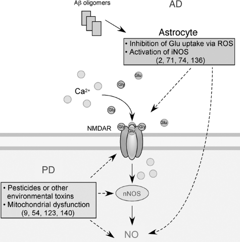
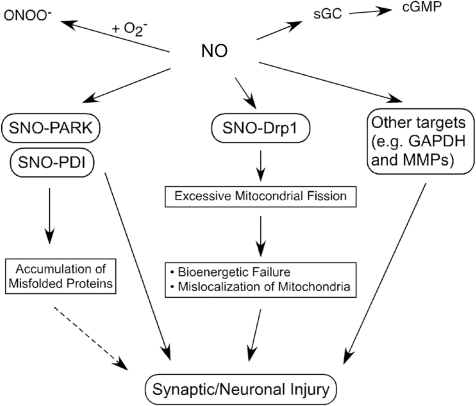
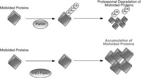
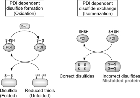
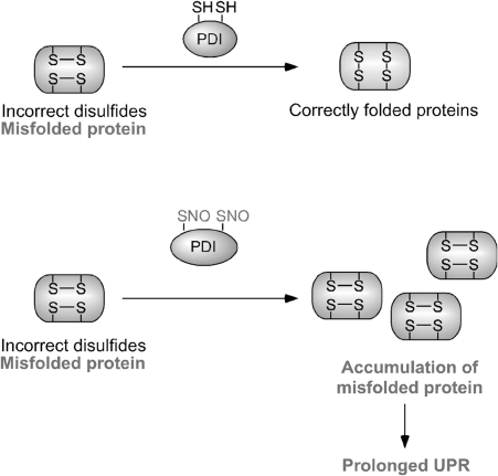
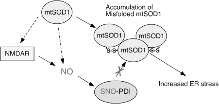
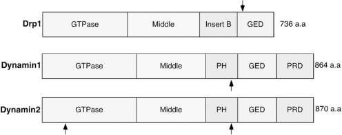
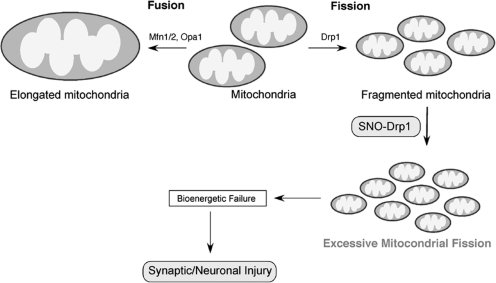
Excessive nitrosative and oxidative stress is thought to trigger cellular signaling pathways leading to neurodegenerative conditions. Such redox dysregulation can result from many cellular events, including hyperactivation of the N-methyl-D-aspartate-type glutamate receptor, mitochondrial dysfunction, and cellular aging. Recently, we and our colleagues have shown that excessive generation of free radicals and related molecules, in particular nitric oxide species (NO), can trigger pathological production of misfolded proteins, abnormal mitochondrial dynamics (comprised of mitochondrial fission and fusion events), and apoptotic pathways in neuronal cells. Emerging evidence suggests that excessive NO production can contribute to these pathological processes, specifically by S-nitrosylation of specific target proteins. Here, we highlight examples of S-nitrosylated proteins that regulate misfolded protein accumulation and mitochondrial dynamics. For instance, in models of Parkinson's disease, these S-nitrosylation targets include parkin, a ubiquitin E3 ligase and neuroprotective molecule, and protein-disulfide isomerase, a chaperone enzyme for nascent protein folding. S-Nitrosylation of protein-disulfide isomerase may also be associated with mutant Cu/Zn superoxide dismutase toxicity in amyotrophic lateral sclerosis. Additionally, in models of Alzheimer's disease, excessive NO generation leads to the formation of S-nitrosylated dynamin-related protein 1 (forming SNO-Drp1), which contributes to abnormal mitochondrial fragmentation and resultant synaptic damage.


Figures

FIG. 1.
FIG. 1.
Possible mechanisms whereby Ca2+ signaling contributes to NO generation in neurodegenerative conditions. Hyperactivation of N-methyl-d-aspartate receptors (NMDARs) by glutamate (Glu) and glycine (Gly) induces excessive Ca2+ influx and activation of neuronal nitric oxide synthase (nNOS). nNOS produces NO from l-arginine. In Alzheimer's disease (AD), soluble oligomers of amyloid-β (Aβ) peptide, thought to be a key mediator in AD pathogenesis, can facilitate neuronal NO production in both NMDAR-dependent and NMDAR-independent manners (2, 71, 74, 136). In Parkinson's disease (PD), mitochondrial dysfunction caused by pesticides, herbicides, or other environmental toxins can trigger NO production, possibly via mitochondrial pathways and the NMDAR/Ca2+ cascade (9, 54, 123, 140). Note that in addition to reactive nitrogen species, reactive oxygen species (ROS) are also produced in response to Aβ and pesticides.
FIG. 2.
FIG. 2.
Overproduction of NO triggers formation of S-nitrosylated (SNO) proteins. NO produced by nNOS reacts with sulfhydryl groups to form SNO proteins. Physiological levels of NO mediate neuroprotective effects, at least in part, by S-nitrosylating the NMDAR and caspases, thus inhibiting their activity. In contrast, we postulate that overproduction of NO can be neurotoxic via S-nitrosylation of Parkin (forming SNO-PARK), protein-disulfide isomerase (PDI) (forming SNO-PDI), GAPDH, MMP-2/9, PrxII, and COX-2. S-Nitrosylated parkin and PDI contribute to neuronal cell injury by triggering accumulation of misfolded proteins. S-Nitrosylation of dynamin-related protein 1 (Drp1) (forming SNO-Drp1) causes excessive mitochondrial fragmentation in neurodegenerative conditions. NO also activates soluble guanylate cyclase (sGC) to produce cyclic guanosine-3′,5′-monophosphate (cGMP), and cGMP can activate cGMP-dependent protein kinase. Peroxynitrite (ONOO), derived from reaction of NO and superoxide anion (O2•−), can oxidize vicinal sulfhydryl groups to disulfide bonds and can also nitrate tyrosine residues to form 3-nitrotyrosine.
FIG. 3.
FIG. 3.
Possible mechanism whereby S-nitrosylated parkin contributes to the accumulation of aberrant proteins and neuronal damage. The ubiquitin E3 ligase parkin is believed to be involved in ubiquitin-proteasome-dependent degradation of misfolded proteins (top). S-Nitrosylation of parkin alters its E3 ligase activity, thus inducing ubiquitin-proteasome system dysfunction (bottom). Thus, S-nitrosylation of parkin (forming SNO-Parkin) can contribute to neuronal cell injury in part by triggering accumulation of misfolded proteins.
FIG. 4.
FIG. 4.
Molecular mechanisms of PDI-dependent oxidative protein folding in the endoplasmic reticulum (ER). Left: Oxidized PDI (top), which contains a disulfide bond at its active site, catalyzes the formation of a disulfide bond in a substrate protein (bottom), resulting in the reduction of PDI. Conversely, the ER oxidoreduction protein, Ero1, can reoxidize and regenerate the PDI active site. By repeating this cycle, PDI can continuously insert disulfide bonds into different substrate proteins. Right: In the early process of protein folding in the ER, cysteine residues often form inaccurate disulfide bonds (resulting in a misfolded protein). The isomerase activity of PDI (top) converts these incorrect disulfide bonds to their correct native form. This reaction occurs through breakage of substrate disulfide, formation of intramolecular disulfide, and reformation of intermolecular disulfide bonds with different thiols in the target substrate protein (bottom). For simplicity, the redox state of only one PDI active site is shown.
FIG. 5.
FIG. 5.
Possible mechanism whereby S-nitrosylated PDI contributes to the accumulation of aberrant proteins and neuronal damage. PDI is known to associate with nascent or misfolded proteins and to refold them correctly (top). Under conditions of nitrosative stress, the decreased chaperone and isomerase activity of PDI due to S-nitrosylation enhances accumulation of misfolded proteins, thus activating a prolonged unfolded protein response (UPR) and neuronal cell death (bottom). In this manner, S-nitrosylation of PDI (forming SNO-PDI) can contribute to neuronal cell injury, in part by triggering accumulation of misfolded proteins.
FIG. 6.
FIG. 6.
Possible mechanism whereby S-nitrosylated PDI contributes to the accumulation of aberrant mutant Cu,Zn superoxide dismutase (mtSOD1) and neuronal damage in familial amyotrophic lateral sclerosis. Amyotrophic lateral sclerosis-linked mtSOD1 is prone to form high-molecular-weight complexes (or aggregates) that contain non-native disulfide bonds. Accumulation of mtSOD1 is known to trigger ER stress. Although most mtSOD1 is present in the cytoplasm, recent lines of evidence suggest that mtSOD1 may be secreted via the ER-Golgi pathway. Further, the ER resident protein PDI binds to both wild-type SOD1 and mtSOD1, thereby reducing mtSOD1 aggregation and toxicity (5, 127). Since S-nitrosylation inhibits the enzymatic activity of PDI, overproduction of NO may contribute to mtSOD1 toxicity via the formation of SNO-PDI. Excessive NMDAR activity may contribute to mtSOD1 toxicity via production of NO to facilitate SNO-PDI formation (130).
FIG. 7.
FIG. 7.
Schematic structure of the domains of Drp1 and Dynamin1/2. The length and topology of amino acids for each region are shown. Each protein has a GTPase domain, a middle domain, and a GTPase effector domain (GED). Dynamin1/2 also contains a pleckstrin homology (PH) domain and a highly basic C-terminal proline-rich domain (PRD). Arrows indicate S-nitrosylated cysteine residues (Cys644 of Drp1, Cys 607 of Dynamin1, and Cys 60 and 607 of Dynamin2). S-Nitrosylation of these proteins upregulates their GTPase activity (29, 64, 128).
FIG. 8.
FIG. 8.
Possible mechanism whereby S-nitrosylated Drp1 contributes to abnormal mitochondrial function and neuronal damage. In healthy neurons, mitochondria continuously undergo fission and fusion to maintain their integrity and insure their presence at critical locations. Mitochondrial fission is driven by fission proteins such as Drp1 and fis1. Components involved in mitochondrial membrane fusion include Opa1 and mitofusin (Mfn)1/2. The NO group reacts with a critical cysteine thiol on Drp1 by S-nitrosylation to form SNO-Drp1. SNO-Drp1 contributes to synaptic and neuronal injury by leading to excessive mitochondrial fission/fragmentation and bioenergetic impairment.

References

    1. Abu-Soud HM. Stuehr DJ. Nitric oxide synthases reveal a role for calmodulin in controlling electron transfer. Proc Natl Acad Sci U S A. 1993;90:10769–10772. - PMC - PubMed
    1. Akama KT. Van Eldik LJ. Beta-amyloid stimulation of inducible nitric-oxide synthase in astrocytes is interleukin-1beta- and tumor necrosis factor-alpha (TNFalpha)-dependent, and involves a TNFalpha receptor-associated factor- and NFkappaB-inducing kinase-dependent signaling mechanism. J Biol Chem. 2000;275:7918–7924. - PubMed
    1. Ankarcrona M. Dypbukt JM. Bonfoco E. Zhivotovsky B. Orrenius S. Lipton SA. Nicotera P. Glutamate-induced neuronal death: a succession of necrosis or apoptosis depending on mitochondrial function. Neuron. 1995;15:961–973. - PubMed
    1. Arnesano F. Banci L. Bertini I. Martinelli M. Furukawa Y. O'Halloran TV. The unusually stable quaternary structure of human Cu,Zn-superoxide dismutase 1 is controlled by both metal occupancy and disulfide status. J Biol Chem. 2004;279:47998–48003. - PubMed
    1. Atkin JD. Farg MA. Turner BJ. Tomas D. Lysaght JA. Nunan J. Rembach A. Nagley P. Beart PM. Cheema SS. Horne MK. Induction of the unfolded protein response in familial amyotrophic lateral sclerosis and association of protein-disulfide isomerase with superoxide dismutase 1. J Biol Chem. 2006;281:30152–30165. - PubMed

MeSH terms