

doi: 10.1042/BJ20100847.
Chondroitin sulfate N-acetylgalactosaminyltransferase-1 is required for normal cartilage development
Affiliations
- PMID: 20812917
- PMCID: PMC2995422
- DOI: 10.1042/BJ20100847
Item in Clipboard
Chondroitin sulfate N-acetylgalactosaminyltransferase-1 is required for normal cartilage development
Biochem J.
.
Abstract
CS (chondroitin sulfate) is a glycosaminoglycan species that is widely distributed in the extracellular matrix. To understand the physiological roles of enzymes involved in CS synthesis, we produced CSGalNAcT1 (CS N-acetylgalactosaminyltransferase 1)-null mice. CS production was reduced by approximately half in CSGalNAcT1-null mice, and the amount of short-chain CS was also reduced. Moreover, the cartilage of the null mice was significantly smaller than that of wild-type mice. Additionally, type-II collagen fibres in developing cartilage were abnormally aggregated and disarranged in the homozygous mutant mice. These results suggest that CSGalNAcT1 is required for normal CS production in developing cartilage.
Figures
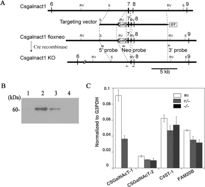
(A) Construct of the targeting vector for producing the CSGalNAcT1-null mice. The numbers represent the exon number (exon 1 is defined as the first exon of this gene where the transcription starts). RV, EcoRV site; S, ScaI site. (B) Immunoblot analysis using an anti-CSGalNAcT1 antibody to probe head homogenate from wild-type mice (lane 2), heterozygous (CSGalNAcT1+/−) mice (lane 3) and null mice (CSGalNAcT1−/−) at E18.5. Lane 1 shows the negative control (wild-type homogenate after absorption by the recombinant CSGalNAcT1 protein). Note that no reactivity was evident in the homogenate from null mice (lane 4). (C) RT–PCR analysis of genes encoding several enzymes involved in CS synthesis. The expression level is shown as a ratio of each gene to G3pdh for normalization. Fam20B is an enzyme that phosphorylates xylose [23]. Note that no other specific enzymes showed elevated expression to compensate for the loss of CSGalNAcT1 protein.
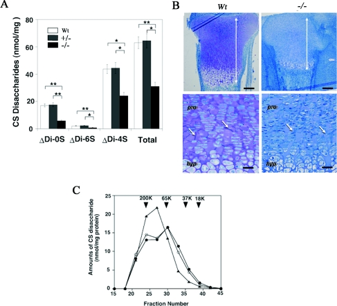
(A) The total amount and disaccharide analysis of the cartilage in wild-type (Wt), heterozygous (+/−; CSGalNAcT1+/−) and null (−/−; CSGalNAcT1−/−) mice. Student's t test was performed to compare the disaccharide amount derived from CS in the null mice with that in the wild-type or the heterozygous mice. Results are shown as mean±S.E.M. *P<0.05; **P<0.01 (n=6). Δdi-0S, ΔHexAα1-3GalNAc; Δdi-4S, ΔHexAα1-3GalNAc(4S); Δdi-6S, HexAα1-3GalNAc(6S). (B) Toluidine Blue staining of the epiphyseal cartilage from E18.5 wild-type (Wt; left) and CSGalNAcT1-null (−/−) fetuses. Metachromasia of Toluidine Blue (purple colour) can be seen in the wild-type cartilage, whereas no metachromasia is discernible in the null mice cartilage. Note the reduced size of the epiphyseal cartilage in the null mice, compared with their wild-type counterpart (bidirectional arrows, upper panels). The extracellular spaces in the cartilage of null mice have many spicules (arrows, lower panels), whereas the intercellular regions from wild-type mice do not. pro, proliferative zone; hyp, hypertrophic zone. Scale bars: in upper panels, 200 μm; in lower panels, 50 μm. (C) Gel-filtration analysis of the length of CS sugar chains in the E18.5 cartilage in wild-type (●), heterozygous (CSGalNAcT1+/−; ○) and null (CSGalNAcT1−/−; ▲) mice. There are no significant differences in the total amount of CS loaded on to the gels among groups. Note that the second peak between fraction numbers 30 and 35 is present both in wild-type and heterozygous mice, but not in CSGalNAcT1-null mice, indicating that the size of the GAG chains of CS changed in CSGalNAcT1-null mice. Arrowheads indicate the size of the molecular-mass-marker standards [mean molecular masses (K, kDa): 200, 65.5, 37.5 and 18.1 respectively; all from Sigma]. The calibration of the Superdex 200 column was performed using a series of size-defined commercial dextran polysaccharides. The results shown represent one of three series of independent experiments, where the three series of experiments gave essentially identical results.
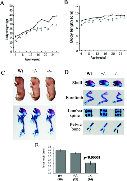
(A and B) The body weight (A) and the body length (B) in the wild-type (●) and CSGalNAcT1-null mice (○) during postnatal development. At 4 weeks after birth, the body mass and the body length of the null mice were slightly reduced compared with those of the wild-type.*P<0.05 (Student's t test). (C) The fetal body (top panel) and skeleton (bottom panel) of wild-type (Wt), heterozygous (+/−) and null (−/−) mice. (D) Various bone segments of the fetal mice. Note that only forelimb bones of the null mice are shorter than those of wild-type mice. (E) Measurements of femur length. The femurs at E18.5 of the null mice are significantly shorter than those of wild-type and heterozygous mice (Student's t test; P<0.00001). The number of the femurs measured for each genotype is shown in parentheses. The results are shown as means±S.E.M.
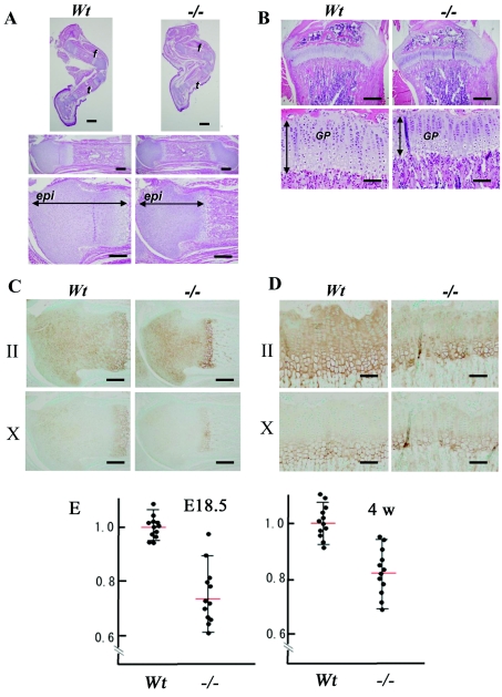
(A) Histological views of the epiphyses of E18.5 wild-type (Wt) and CSGalNAcT1-null (−/−) mice. Upper panels: hind limbs (f, femur; t, tibia), middle panels: the femurs of these littermates; lower panels: higher magnification views of the middle panels focusing on the epiphyseal cartilage. Consistent with the decreased size of femurs and tibiae of the null mice, the femoral epiphyseal cartilage (epi) was reduced in size in null mice compared with that of their wild-type counterparts. Scale bar: upper panels, 1 mm; middle panels, 500 μm; lower panels, 300 μm. (B) Histological findings on the growth plates of 4-week-old wild-type (Wt) and null (−/−) mice. Upper and lower panels show lower and higher magnification respectively. The longitudinal length of the growth plates (GP) was shortened in the null mice (bidirectional arrows, lower panels). Scale bars: upper panels, 800 μm; lower panels, 200 μm. (C and D) Immunodetection of type-II (II; upper panels) and type-X (X; lower panels) collagen in the epiphyses of the wild-type (Wt) and null (−/−) E18.5 fetuses (C) and 4-week-old mice (D). In (C), the type-II collagen-positive area (brown) was seen throughout the epiphyses of both Wt and null mice, despite the finding that cartilage area of the null mice was reduced (upper panels). Even in (D), the reduced size of the type-II collagen reactive-growth plate in the null mice was observed. Judging from the hypertrophic zone marker type-X collagen immunostaining, cartilage from E18.5 (C) and four-week-old (D) animals had a reduced hypertrophic zone. Scale bars: (C), 300 μm; (D), 100 μm. (E) Quantitative measurement of the epiphyseal cartilage in area. Both E18.5 and 4-week-old (4w) femurs were collected. (Mann–Whitney U test; P<0.05). The results are shown as means±S.E.M.
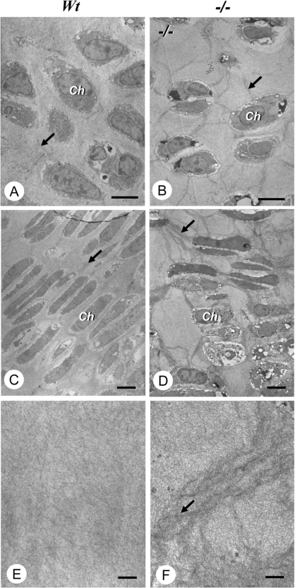
(A, C and E) Wild-type (Wt) mice; (B, D and F) CSGalNAcT1-null (−/−) mice; at E18.5. Micrographs (A) and (B) were obtained from the resting zone, and (C) and (D) from the proliferative zones. Note that many electron-dense extracellular fibrils (arrows) are seen connecting chondrocytes in the resting (B) and proliferative (D) zones of the epiphysis from null mice, whereas only a few such fibrils are observed in both zones (A; resting zone, and C; proliferative zone) of wild-type mice. At a higher magnification, the wild-type cartilage matrix had collagen fibrils that spread radially (E), whereas cartilage of the null mice had a fine meshwork of twisted cartilaginous fibrils (F). Ch, chondrocytes. Scale bars: (A)–(D), 10 μm; (E)–(F), 1 μm.
Similar articles
-
Chondroitin sulfate N-acetylgalactosaminyltransferase 1 is necessary for normal endochondral ossification and aggrecan metabolism.J Biol Chem. 2011 Feb 18;286(7):5803-12. doi: 10.1074/jbc.M110.159244. Epub 2010 Dec 10. J Biol Chem. 2011. PMID: 21148564 Free PMC article.
-
GlcUAβ1-3Galβ1-3Galβ1-4Xyl(2-O-phosphate) is the preferred substrate for chondroitin N-acetylgalactosaminyltransferase-1.J Biol Chem. 2015 Feb 27;290(9):5438-48. doi: 10.1074/jbc.M114.603266. Epub 2015 Jan 7. J Biol Chem. 2015. PMID: 25568321 Free PMC article.
-
Chondroitin sulfate N-acetylgalactosyltransferase-1 knockout shows milder phenotype in experimental autoimmune encephalomyelitis than in wild type.Glycobiology. 2021 Apr 1;31(3):260-265. doi: 10.1093/glycob/cwaa072. Glycobiology. 2021. PMID: 32839819
-
Roles of CSGalNAcT1, a key enzyme in regulation of CS synthesis, in neuronal regeneration and plasticity.Neurochem Int. 2018 Oct;119:77-83. doi: 10.1016/j.neuint.2017.10.001. Epub 2017 Oct 5. Neurochem Int. 2018. PMID: 28987564 Review.
-
The roles of chondroitin-4-sulfotransferase-1 in development and disease.Prog Mol Biol Transl Sci. 2010;93:113-32. doi: 10.1016/S1877-1173(10)93006-8. Prog Mol Biol Transl Sci. 2010. PMID: 20807643 Review.
Cited by
-
Reconsideration of the Semaphorin-3A Binding Motif Found in Chondroitin Sulfate Using Galnac4s-6st-Knockout Mice.Biomolecules. 2020 Oct 30;10(11):1499. doi: 10.3390/biom10111499. Biomolecules. 2020. PMID: 33143303 Free PMC article.
-
Human genetic disorders and knockout mice deficient in glycosaminoglycan.Biomed Res Int. 2014;2014:495764. doi: 10.1155/2014/495764. Epub 2014 Jul 13. Biomed Res Int. 2014. PMID: 25126564 Free PMC article. Review.
-
Chondroitin sulfate-E mediates estrogen-induced osteoanabolism.Sci Rep. 2015 Mar 11;5:8994. doi: 10.1038/srep08994. Sci Rep. 2015. PMID: 25759206 Free PMC article.
-
The different roles of aggrecan interaction domains.J Histochem Cytochem. 2012 Dec;60(12):987-96. doi: 10.1369/0022155412464376. Epub 2012 Sep 26. J Histochem Cytochem. 2012. PMID: 23019016 Free PMC article. Review.
-
Comparison of the effects of exercise with chondroitin sulfate on knee osteoarthritis in rabbits.J Orthop Surg Res. 2018 Jan 22;13(1):16. doi: 10.1186/s13018-018-0722-4. J Orthop Surg Res. 2018. PMID: 29357891 Free PMC article.
References
-
- Gandhi N. S., Mancera R. L. The structure of glycosaminoglycans and their interactions with proteins. Chem. Biol. Drug Des. 2008;72:455–492. - PubMed
-
- Kitagawa H., Uyama T., Sugahara K. Molecular cloning and expression of a human chondroitin synthase. J. Biol. Chem. 2001;276:38721–38726. - PubMed
-
- Kitagawa H., Izumikawa T., Uyama T., Sugahara K. Molecular cloning of a chondroitin polymerizing factor that cooperates with chondroitin synthase for chondroitin polymerization. J. Biol. Chem. 2003;278:23666–23671. - PubMed
-
- Uyama T., Kitagawa H., Tamura J., Sugahara K. Molecular cloning and expression of human chondroitin N-acetylgalactosaminyltransferase: the key enzyme for chain initiation and elongation of chondroitin/dermatan sulfate on the protein linkage region tetrasaccharide shared by heparin/heparan sulfate. J. Biol. Chem. 2002;277:8841–8846. - PubMed
-
- Uyama T., Kitagawa H., Tanaka J., Tamura J., Ogawa T., Sugahara K. Molecular cloning and expression of a second chondroitin N-acetylgalactosaminyltransferase involved in the initiation and elongation of chondroitin/dermatan sulfate. J. Biol. Chem. 2003;278:3072–3078. - PubMed
Publication types
MeSH terms
Substances
LinkOut - more resources
Full Text Sources
Other Literature Sources
Molecular Biology Databases