

Alcohol and the prefrontal cortex
- PMID: 20813246
- PMCID: PMC3593065
- DOI: 10.1016/S0074-7742(10)91009-X
Alcohol and the prefrontal cortex
Abstract
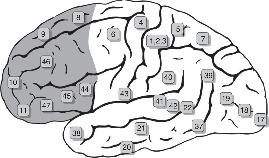
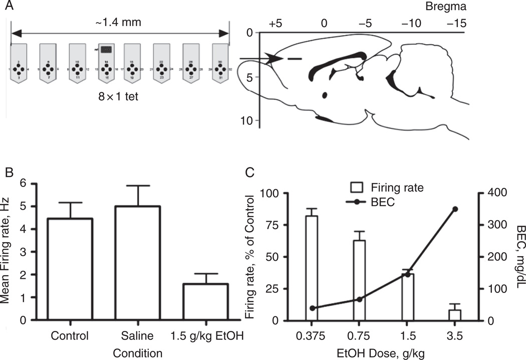
The prefrontal cortex occupies the anterior portion of the frontal lobes and is thought to be one of the most complex anatomical and functional structures of the mammalian brain. Its major role is to integrate and interpret inputs from cortical and sub-cortical structures and use this information to develop purposeful responses that reflect both present and future circumstances. This includes both action-oriented sequences involved in obtaining rewards and inhibition of behaviors that pose undue risk or harm to the individual. Given the central role in initiating and regulating these often complex cognitive and behavioral responses, it is no surprise that alcohol has profound effects on the function of the prefrontal cortex. In this chapter, we review the basic anatomy and physiology of the prefrontal cortex and discuss what is known about the actions of alcohol on the function of this brain region. This includes a review of both the human and animal literature including information on the electrophysiological and behavioral effects that follow acute and chronic exposure to alcohol. The chapter concludes with a discussion of unanswered questions and areas needing further investigation.
Copyright 2010 Elsevier Inc. All rights reserved.
Figures
References
-
- Adams KM, Gilman S, Koeppe RA, Kluin KJ, Brunberg JA, Dede D, Berent S, Kroll PD. Neuropsychological deficits are correlated with frontal hypometabolism in positron emission tomography studies of older alcoholic patients. Alcohol. Clin. Exp. Res. 1993;17:205–210. - PubMed
-
- Alexander-Kaufman K, James G, Sheedy D, Harper C, Matsumoto I. Differential protein expression in the prefrontal white matter of human alcoholics: A proteomics study. Mol. Psychiatry. 2006;11:56–65. - PubMed
-
- Araneda R, Andrade R. 5-Hydroxytryptamine2 and 5-hydroxytryptamine 1A receptors mediate opposing responses on membrane excitability in rat association cortex. Neuroscience. 1991;40:399–412. - PubMed
-
- Arbib MA. Schemas for the temporal organization of behaviour. Hum. Neurobiol. 1985;4:63–72. - PubMed
Publication types
MeSH terms
Substances
Grants and funding
LinkOut - more resources
Full Text Sources
