. 2010 Sep 3;142(5):699-713.
doi: 10.1016/j.cell.2010.07.044.

Anti-CD47 antibody synergizes with rituximab to promote phagocytosis and eradicate non-Hodgkin lymphoma

Affiliations

Anti-CD47 antibody synergizes with rituximab to promote phagocytosis and eradicate non-Hodgkin lymphoma

Mark P Chao et al. Cell. .

Abstract

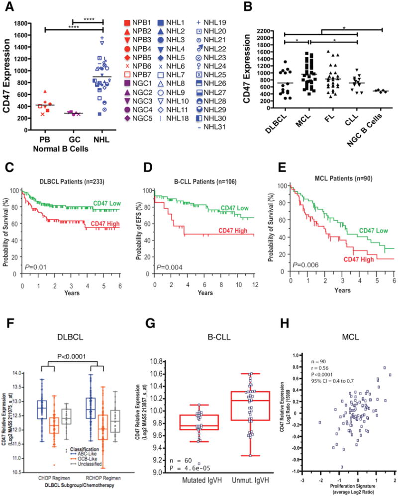
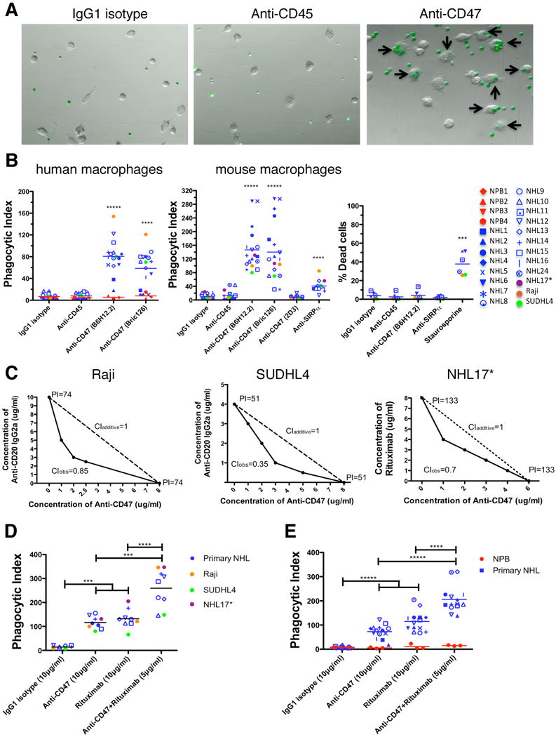
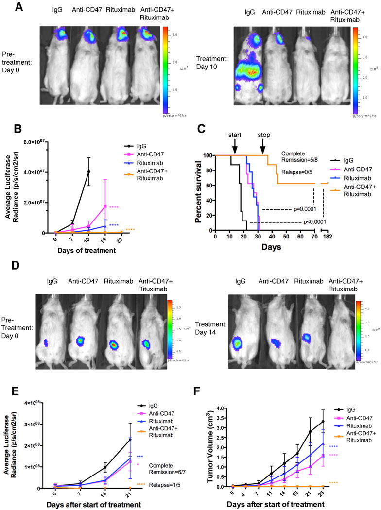
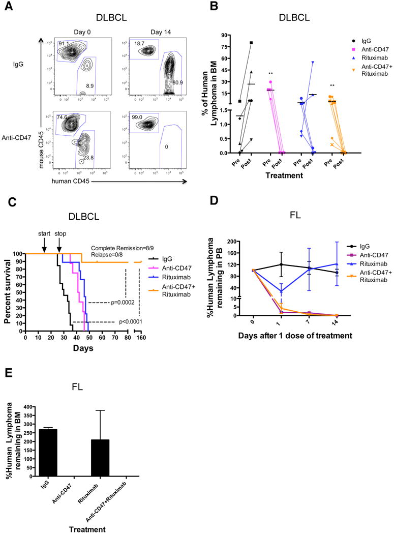
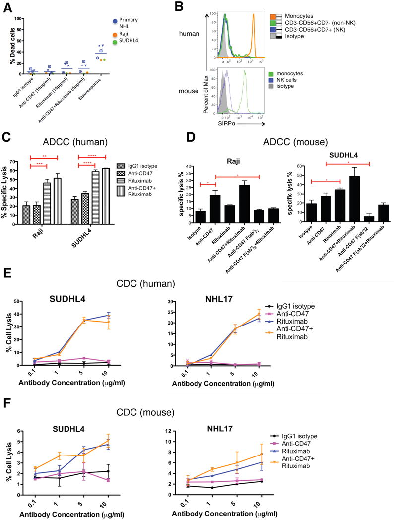
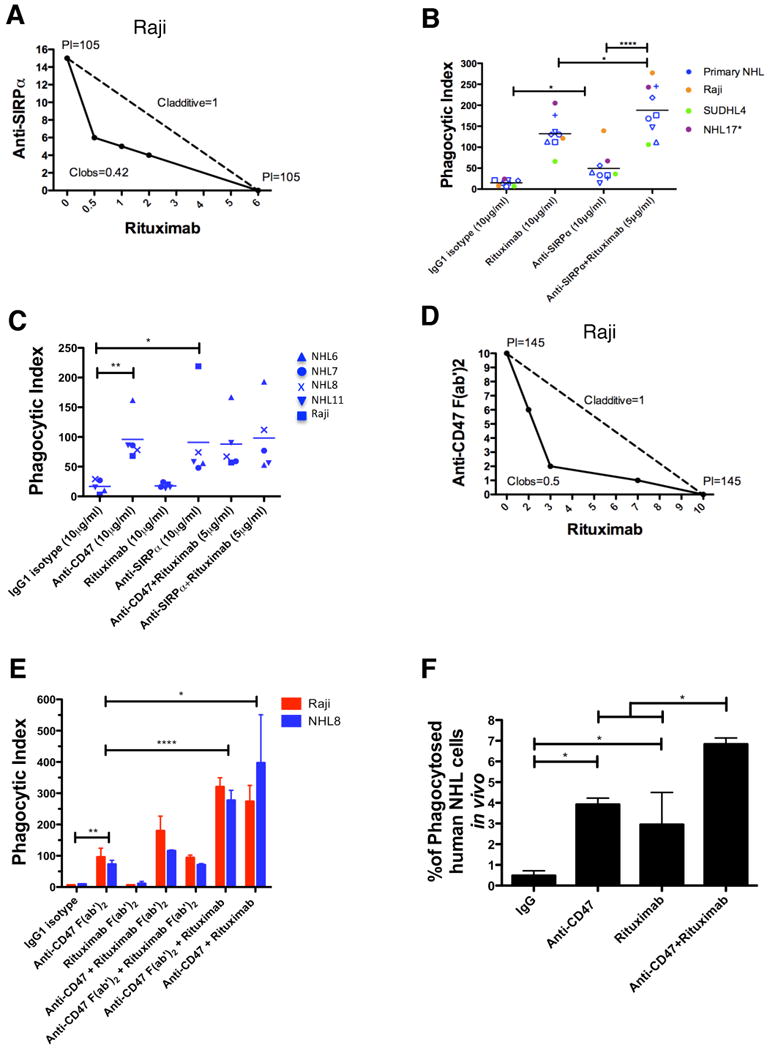
Monoclonal antibodies are standard therapeutics for several cancers including the anti-CD20 antibody rituximab for B cell non-Hodgkin lymphoma (NHL). Rituximab and other antibodies are not curative and must be combined with cytotoxic chemotherapy for clinical benefit. Here we report the eradication of human NHL solely with a monoclonal antibody therapy combining rituximab with a blocking anti-CD47 antibody. We identified increased expression of CD47 on human NHL cells and determined that higher CD47 expression independently predicted adverse clinical outcomes in multiple NHL subtypes. Blocking anti-CD47 antibodies preferentially enabled phagocytosis of NHL cells and synergized with rituximab. Treatment of human NHL-engrafted mice with anti-CD47 antibody reduced lymphoma burden and improved survival, while combination treatment with rituximab led to elimination of lymphoma and cure. These antibodies synergized through a mechanism combining Fc receptor (FcR)-dependent and FcR-independent stimulation of phagocytosis that might be applicable to many other cancers.


Figures

Figure 1
Figure 1. CD47 Expression is Increased on NHL Cells Compared to Normal B Cells, Confers a Worse Clinical Prognosis, and Correlates with Adverse Molecular Features in Multiple NHL Subtypes
(A) CD47 expression on normal peripheral blood (PB) B cells (CD19+), normal germinal center (GC) B cells (CD3-CD5-CD20+CD10+CD38+), and NHL B cells (CD19+) was determined by flow cytometry, and mean fluorescence intensity was normalized for cell size. Each point represents a different patient sample: DLBCL=2, CLL=15, MCL=4, FL=6, MZL=2, and pre-B ALL=1 (****p<0.0001). Normalized mean expression (and range) for each population were: normal PB B cells 420.9 (267.3-654.0), normal GC B cells 482.5 (441.1-519.9), and NHL 888.5 (270.1-1553). (B) CD47 expression across NHL subtypes including DLBCL (DL, n=15), MCL (n=34), FL (n=28), and B-CLL (n=14) was determined as in A. Normalized mean expression (and range) for each population were: DL 725.7 (261.2 – 1344), MCL 1055 (444.2-2196), FL 825.1 (283.6-1546), CLL 713.6 (432.8-1086), (*p<0.05). (B to D) Prognostic influence of CD47 mRNA expression is shown on overall (C and E) and event-free (D) survival of patients with DLBCL, B-CLL, and MCL. For DLBCL and CLL, stratification into low and high CD47 expression groups was based on an optimal threshold determined by microarray analysis; this cut point was internally validated for both DLBCL and CLL, and also externally validated in an independent DLBCL cohort. For MCL, stratification relative to the median was employed as an optimal cut point could not be defined. Significance measures are based on log-likelihood estimates of the p-value, when treating CD47 expression as a continuous variable, with corresponding dichotomous indices also provided in Table S1. (F to H) CD47 mRNA expression is shown in relation to cell-of-origin classification for DLBCL (F), immunoglobulin heavy chain mutation status (IgVH) for CLL (G), and proliferation index for MCL (H). Analyses for C-H employed publicly available datasets for NHL patients (Table S1). NGC=normal germinal center, ABC=activated B cell-like, GCB=germinal center B cell-like. See also Figure S1 and Table S1.
Figure 2
Figure 2. Blocking Antibodies Against CD47 Enable Phagocytosis of NHL Cells by Macrophages and Synergize with Rituximab in Vitro
(A) CFSE-labeled NHL cells were incubated with human macrophages and the indicated antibodies and examined by immunofluorescence microscopy to detect phagocytosis (arrows). Photomicrographs from a representative NHL sample are shown. (B) Phagocytic indices of primary human NHL cells (blue), normal peripheral blood (NPB) cells (red), and NHL cell lines (purple, orange, and green) were determined using human (left) and mouse (middle) macrophages. Antibody-induced apoptosis (right panel) was tested by incubating NHL cells with the indicated antibodies or staurosporine without macrophages, and assessing the percentage of apoptotic and dead cells (% annexin V and/or PI positive). (C) Synergistic phagocytosis by anti-CD47 antibody (B6H12.2) and anti-CD20 mAbs was examined by isobologram analysis and determination of combination indices (CI). CIobs indicates observed results, and the dashed line indicates the expected results if antibodies were additive. (D,E) Synergy between anti-CD47 antibody and rituximab in the phagocytosis of NHL and NPB cells was assessed by determining the phagocytic index when incubated with a combination of both antibodies compared to either antibody alone at twice the dose, with either mouse (D) or human (E) macrophages. NHL17*: cell line derived from primary sample NHL17. ***p<0.001, ****p<0.0001, *****p<0.00001. Figure 2B p-values represent comparison against IgG1 isotype control antibody. See also Figure S2.
Figure 3
Figure 3. Ex Vivo Coating of NHL Cells with an Anti-CD47 Antibody Inhibits Tumor Engraftment
(A-F) Luciferase-expressing Raji (A) and SUDHL4 (C) cells were assessed for ex vivo antibody coating by flow cytometry. SCID mice transplanted with Raji (B) and SUDHL4 (D) were subject to bioluminescent imaging (1-IgG1 isotype control, 2-anti-CD45, 3 and 4-anti-CD47, 5-luciferase negative control). Bioluminescence for Raji (E) and SUDHL4 (F) engrafted mice was quantified (n=3 per antibody condition). No tumor engraftment was observed in mice transplanted with anti-CD47-coated cells compared to IgG-coated cells (p<0.05) for both Raji and SUDHL4, as assessed by bioluminescent imaging. Data are represented as mean +/- SD. (G) Bulk lymphoma cells from a human NHL patient were assessed for ex vivo antibody coating by flow cytometry. (H) Compared to IgG1 isotype control, anti-CD47 antibody pre-treatment inhibited engraftment of NHL cells (p<0.0001) while anti-CD45 coated cells engrafted similarly to controls (p=0.54). All p-values were determined using Fisher's exact test. See also Figure S3.
Figure 4
Figure 4. Combination Therapy with anti-CD47 Antibody and Rituximab Eliminates Lymphoma in both Disseminated and Localized Human NHL Xenotransplant Mouse Models
(A) NSG mice transplanted intravenously with luciferase-expressing Raji cells were treated with the indicated antibodies (n=8 per treatment group). Luciferase imaging of representative mice from pre- and 10 days post-treatment are shown (A) and averaged for all mice in each treatment group (B). (C) Kaplan-Meier survival analysis was performed (Table S2). p-values compare IgG control to combination antibody treatment or anti-CD47 antibody/rituximab single antibody to combination. Arrows indicate start (day 14) and stop (day 35) of treatment. (D) Luciferase-expressing Raji cells were transplanted subcutaneously in the flank of NSG mice. When palpable tumors (∼0.1cm3) formed, treatment began with the indicated antibodies. Luciferase imaging of representative mice from each treatment group is shown before (day 0) and during (day 14) treatment. (E) Quantified bioluminescence was determined and averaged for all mice in each treatment group (n=7). (F) Tumor volume was measured with average volume shown. p-values were derived from a two-way ANOVA and compared to IgG treatment. *p<0.05, ***p<0.001, ****p<0.0001. Complete remission was defined as the number of mice with no evidence of tumor at the indicated date. Relapse was defined as the number of mice achieving a complete remission that later developed recurrence of tumor growth. For panel E, one mouse that achieved a complete remission died of non-tumor related causes. Data presented in B, E, and F are mean values +/- SD. See also Figure S4 and Table S2.
Figure 5
Figure 5. Combination Therapy with Anti-CD47 Antibody and Rituximab Eliminates Lymphoma in Primary Human NHL Xenotransplant Mouse Models
(A,B) Sublethally-irradiated NSG mice were transplanted intravenously with cells from a primary DLBCL patient (NHL7) and treated with the indicated antibodies. Flow cytometry of human lymphoma engraftment in the bone marrow of two representative mice is shown pre- and 14 days post-antibody treatment in (A). Data from all mice is included in (B). **p<0.01, comparing pre- and post-treatment values for each respective antibody treatment. (C) Kaplan-Meier survival analysis (Table S3) of DLBCL-engrafted mice from each antibody treatment cohort is shown (n≥10 per antibody group), with p-values calculated comparing control IgG to combination antibody treatment or anti-CD47 antibody/rituximab single antibody to combination treatment. Arrows indicate start (day 14) and stop (day 28) of treatment. (D-E) Mice engrafted intravenously with a primary FL patient sample (NHL31) were treated with a single dose of the indicated antibodies. Compared to IgG control and rituximab, anti-CD47 antibody alone or in combination with rituximab eliminated tumor burden in the peripheral blood (p=0.04, 2-way ANOVA), and bone marrow (p<0.001, t-test). (E) Lyphoma engraftment in the bone marrow was determined 14 days post-treatment. Each antibody treatment group consisted of 3 mice. For mice reported in panels D and E, human lymphoma chimerism was between 5-25% in the peripheral blood and bone marrow. See also Figure S5 and Table S3.
Figure 6
Figure 6. Synergy Between Anti-CD47 Antibody and Rituximab Does Not Occur Through NK Cells or Complement
(A) NHL cells were incubated with the indicated soluble antibodies for 2 hours and the percentage of dead cells was calculated (% Annexin V+ and/or 7-AAD+). No statistically significant difference in % dead cells was observed with the combination of anti-CD47 antibody and rituximab compared to either anti-CD47 antibody alone (p=0.24) or rituximab alone (p=0.95). (B) SIRPα expression is shown for both human and mouse NK cells as determined by flow cytometry. (C,D) Chromium release assays measuring ADCC were performed in triplicate with human (C) and mouse (D) at an effector:target ratio of 17.5:1 and percent specific lysis is reported. Antibodies were incubated at 10μg/ml except anti-CD47 full length or F(ab′)2 antibody+rituximab (5μg/ml). (E) CDC assay with human complement was performed in duplicate. Compared to IgG1 isotype control, anti-CD47 antibody did not enable CDC (p>0.2), while rituximab did (p<0.001) by 2-way ANOVA for both SUDHL4 and NHL17*. Combination treatment with anti-CD47 antibody and rituximab did not enable greater levels of CDC compared to rituximab (p=0.78). (F) CDC assay with mouse complement was performed in duplicate. Compared to IgG1 isotype control, anti-CD47 antibody did not enable CDC (p>0.25) while rituximab did (p=0.03, p=0.08, respectively) for both SUDHL4 and NHL17*. No difference in CDC between CD47 antibody+rituximab and rituximab alone was observed (p>0.13) for both SUDHL4 and NHL17*. *p<0.05, **p<0.01, ***p<0.001, ****p<0.0001. NHL17*=Primary NHL17 cells expanded in culture. See also Figure S6.
Figure 7
Figure 7. Anti-CD47 Antibody Synergizes with Rituximab Through FcR-Independent and FcR-Dependent Mechanisms
(A) Isobologram analysis of phagocytosis induced by anti-SIRPα antibody and rituximab is shown for Raji cells and mouse macrophages. (B,C) NHL cells were incubated in vitro with the indicated antibodies in the presence of wild type (B) or Fcγr-/- (C) mouse macrophages, and the phagocytic index was determined. (D) Isobologram analysis of phagocytosis induced by anti-CD47 F(ab′)2 antibody and rituximab is shown for Raji cells and mouse macrophages. (E) NHL cells were incubated with wild type mouse macrophages in the presence of the indicated full length or F(ab′)2 antibodies (single antibodies at 10μg/ml, combination antibodies at 5μg/ml each) and the phagocytic index was determined. (F) The level of in vivo phagocytosis measured as the percentage of mouse macrophages containing phagocytosed GFP+ Raji cells (hCD45-GFP+F4/80+) was determined by flow cytometry of livers from mice engrafted with GFP+ Raji cells and then treated with the indicated antibodies (see methods), with each treatment group performed in duplicate. Compared to IgG control, anti-CD47 antibody and rituximab enabled increased levels of phagocytosis. Compared to anti-CD47 antibody alone, combination anti-CD47 antibody and rituximab enabled higher levels of phagocytosis. *p<0.05, **p<0.01, ****p<0.0001. See also Figure S7.

References

    1. A predictive model for aggressive non-Hodgkin's lymphoma. The International Non-Hodgkin's Lymphoma Prognostic Factors Project. N Engl J Med. 1993;329:987–994. - PubMed
    1. Adams GP, Weiner LM. Monoclonal antibody therapy of cancer. Nat Biotechnol. 2005;23:1147–1157. - PubMed
    1. Alizadeh AA, Eisen MB, Davis RE, Ma C, Lossos IS, Rosenwald A, Boldrick JC, Sabet H, Tran T, Yu X, et al. Distinct types of diffuse large B-cell lymphoma identified by gene expression profiling. Nature. 2000;403:503–511. - PubMed
    1. Brown EJ, Frazier WA. Integrin-associated protein (CD47) and its ligands. Trends Cell Biol. 2001;11:130–135. - PubMed
    1. Cartron G, Watier H, Golay J, Solal-Celigny P. From the bench to the bedside: ways to improve rituximab efficacy. Blood. 2004;104:2635–2642. - PubMed

Publication types