. 2011 Feb;19(1):84-94.
doi: 10.1109/TNSRE.2010.2065241. Epub 2010 Sep 2.

Effects of biphasic current pulse frequency, amplitude, duration, and interphase gap on eye movement responses to prosthetic electrical stimulation of the vestibular nerve

Affiliations

Effects of biphasic current pulse frequency, amplitude, duration, and interphase gap on eye movement responses to prosthetic electrical stimulation of the vestibular nerve

Natan S Davidovics et al. IEEE Trans Neural Syst Rehabil Eng. 2011 Feb.

Abstract

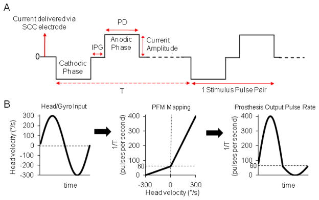
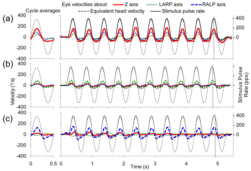
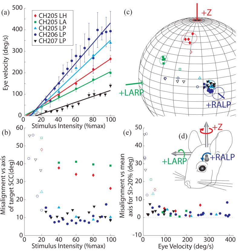
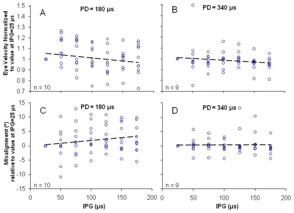
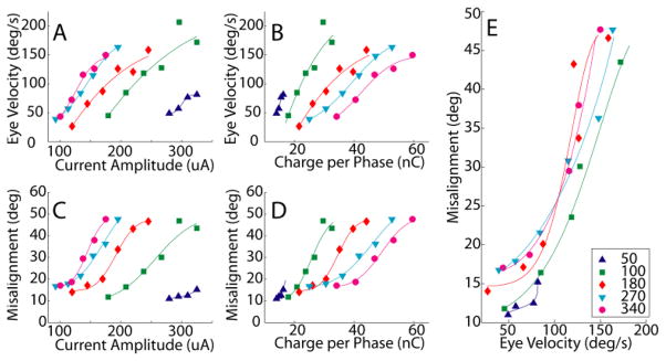
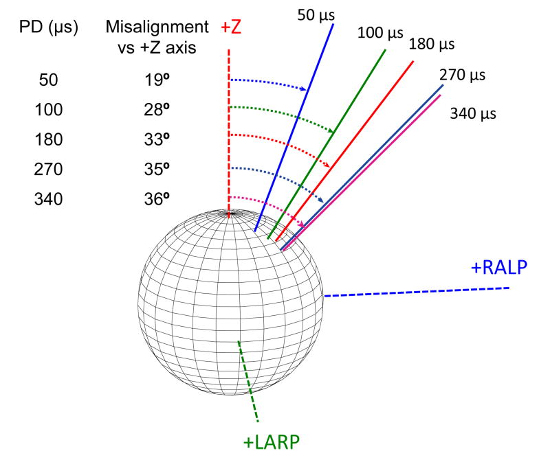
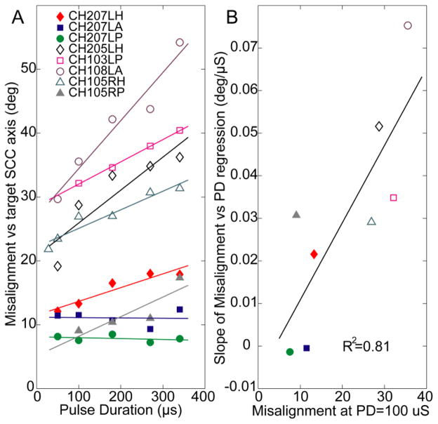
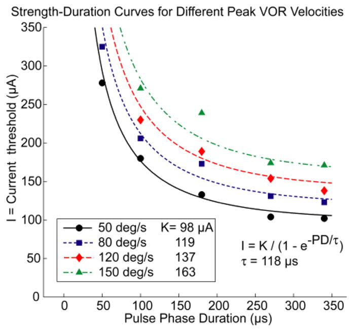
An implantable prosthesis that stimulates vestibular nerve branches to restore sensation of head rotation and vision-stabilizing reflexes could benefit individuals disabled by bilateral loss of vestibular (inner ear balance) function. We developed a prosthesis that partly restores normal function in animals by delivering pulse frequency modulated (PFM) biphasic current pulses via electrodes implanted in semicircular canals. Because the optimal stimulus encoding strategy is not yet known, we investigated effects of varying biphasic current pulse frequency, amplitude, duration, and interphase gap on vestibulo-ocular reflex (VOR) eye movements in chinchillas. Increasing pulse frequency increased response amplitude while maintaining a relatively constant axis of rotation. Increasing pulse amplitude (range 0- 325 μA) also increased response amplitude but spuriously shifted eye movement axis, probably due to current spread beyond the target nerve. Shorter pulse durations (range 28- 340 μs) required less charge to elicit a given response amplitude and caused less axis shift than longer durations. Varying interphase gap (range 25- 175 μs) had no significant effect. While specific values reported herein depend on microanatomy and electrode location in each case, we conclude that PFM with short duration biphasic pulses should form the foundation for further optimization of stimulus encoding strategies for vestibular prostheses intended to restore sensation of head rotation.


Figures

Fig. 1
Fig. 1
(A) Three parameters define a cathodic-first, symmetric, biphasic, electrical stimulation pulse: current amplitude, pulse duration (PD), and interphase gap (IPG). T = time interval between pulse pairs. (B) The stimulus encoding scheme used for each of three dimensions of head rotation converts head velocity about a given semicircular canal axis to modulation of pulse rate f = 1/T on the corresponding electrodes via a piecewise-linear velocity-to-pulse rate mapping meant to efficiently approximate the mean operating characteristic of vestibular nerve afferent fibers. Stimulus intensity = 100% is depicted here.
Fig. 2
Fig. 2
Eye velocity responses to pulse-frequency-modulated, constant current, symmetric biphasic pulses delivered at a modulation frequency of 2 Hz with stimulus intensity = 100% to the left horizontal semicircular (SCC) of CH205 (A), the left anterior SCC of CH207 (B), and the left posterior SCC of CH207 (C). Thick red/solid line indicates the component of eye movement about the horizontal axis, green/dotted line indicates the left anterior-right posterior (LARP) axis, and blue/dashed line indicates the right-anterior/left-posterior (RALP) axis. Thin black/dashed trace shows the angular head velocity about the RALP axis equivalent to the stimulus presented. Thin black/solid trace represents the delivered pulse rate of the stimulus (right-hand ordinate). Vertical gray lines represent the time of peak eye velocity for a given cycle. Mean responses for each dataset are in the left column (SD<19 dps for all datasets). The animal was kept stationary to examine responses to electrical stimulation alone. The largest amplitude trace in each figure represents the eye movement component about the desired axis of rotation while other traces represent undesired eye movement components. Refer to Fig. 3D for orientation of eye movement axes.
Fig. 3
Fig. 3
Peak excitatory response eye velocity (A) and misalignment (B) as a function of stimulus intensity for 5 implanted SCCs. In each case, pulse frequency modulation (PFM) of biphasic current pulses was used to encode a virtual sinusoidal head velocity at 2 Hz as described in text. Stimulus intensity (SI) of 100% corresponds to PFM about a baseline of 60 pps up to 400 pps and down to 0 pps. SI<100% implies a proportional decrease in pulse rate excursion about the baseline. In all cases, VOR response eye velocity increased almost linearly with increasing SI (R2>0.94 for each case), while misalignment between the desired and observed VOR axes remained relatively constant for SI>20%. (C) Axes of responses for each SCC tested cluster tightly in each case about a constant axis for SI>20% (solid symbols) but depart from that axis for lower SI (open symbols). Circles around the data points represent 3 standard deviations of misalignment angle beyond the mean axis of responses for SI>20%. Angle measurement error for each SCC axis (depicted in (D) in reference to an animal head) is based on animal measurements [58]. (E) To more clearly depict the consistency of the misalignment across eye response velocities, misalignment with respect to mean response axes to stimuli with SI>20% for each SCC is plotted against eye velocity. Axis of rotation remains relatively constant for peak excitatory eye responses>50°/s.
Fig. 4
Fig. 4
Effect of variation in interphase gap (IPG). Symmetric biphasic pulses with varying IPG were delivered to 10 implanted SCCs in 5 animals at two different pulse durations (PD), 180 μs and 340 μs. The current amplitude was halfway between threshold and maximum current levels. For the range of IPGs tested, no significant effect was observed on either the VOR response eye velocity (A,B) or axis misalignment (C,D). R2 < 0.05 for regression lines in all panels.
Fig. 5
Fig. 5
Stimulation pulses with varying pulse durations (PD) were delivered to the left horizontal SCC of animal CH207 at a range of current amplitudes. Peak excitatory response eye velocity increased with increasing current (A) and charge per phase (B) for all PD tested. Misalignment also increased with increasing current (C) and charge per phase (D) for all PD tested. Shorter PD stimuli required less charge per phase to evoke eye responses of a given velocity (B) and achieved a given velocity with less change in eye movement axis (E) than did longer PD stimuli. Changes in current (or charge) can encode changes in head velocity below ~75°/s without changing axis, but eye rotation axis shifts dramatically as current is increased to encode higher eye velocities. Curves in A–D are least-mean-square fits of cumulative Gaussian distribution functions; curves in E were derived from those fit to A and C.
Fig. 6
Fig. 6
Longer PD pulses result in responses with greater misalignment versus the desired horizontal axis eye rotation for a given eye velocity of 200°/s (CH205, left horizontal SCC electrode).
Fig. 7
Fig. 7
(A) Misalignment increased with increasing PD for 6 of the 8 SCCs tested; current amplitude chosen in each case to yield half-maximal eye velocity). (B) Cases with the worst (highest) misalignment measured at PD = 100 μs have the greatest potential for improvement with shorter PD (R2 = 0.81).
Fig. 8
Fig. 8
The relationship between PD and current required to elicit eye rotation at a specified peak excitatory response velocity is well described by a family of classic strength-duration curves with time constant 118 μs. [29, 40, 41]

References

    1. Carey JP, Della Santina CC. Principles of applied vestibular physiology. In: Cummings CW, editor. Otolaryngology - Head & Neck Surgery. Elsevier; 2005. pp. 3115–3159.
    1. Black FO, Gianna-Poulin C, Pesznecker SC. Recovery from vestibular ototoxicity. Otology & Neurotology. 2001;22:662–671. - PubMed
    1. Black FO, Pesznecker S, Stallings V. Permanent gentamicin vestibulotoxicity. Otology & Neurotology. 2004;25:559–569. - PubMed
    1. Mamoto Y, Yamamoto K, Imai T, Tamura M, Kubo T. Three-dimensional analysis of human locomotion in normal subjects and patients with vestibular deficiency. Acta Oto-Laryngologica. 2002;122:495–500. - PubMed
    1. Rinne T, Bronstein AM, Rudge P, Gresty MA, Luxon LM. Bilateral loss of vestibular function: clinical findings in 53 patients. Journal of Neurology. 1998;245:314–321. - PubMed

Publication types

MeSH terms