

Modified C-reactive protein might be a target autoantigen of TINU syndrome
- PMID: 20813859
- PMCID: PMC3022254
- DOI: 10.2215/CJN.09051209
Modified C-reactive protein might be a target autoantigen of TINU syndrome
Abstract
Background and objectives: The cross-reactive antigen(s) of tubulointerstitial nephritis and uveitis (TINU) syndrome from renal tubulointerstitia and ocular tissue remain unidentified. The authors' recent study demonstrated that the presence of serum IgG autoantibodies against modified C-reactive protein (mCRP) was closely associated with the intensity of tubulointerstitial lesions in lupus nephritis. The study presented here investigates the possible role of IgG autoantibodies against mCRP in patients with TINU syndrome.
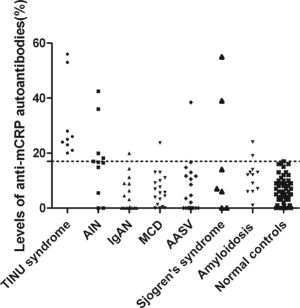
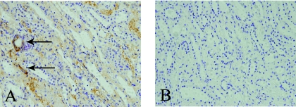
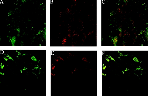
Design, setting, participants, & measurements: mCRP autoantibodies were screened by ELISA with purified human C-reactive protein in 9 patients with TINU syndrome, 11 with drug-associated acute interstitial nephritis, 20 with IgA nephropathy, 19 with minimal change disease, 20 with ANCA-associated vasculitis, 6 with Sjogren's syndrome, and 12 with amyloidosis. mCRP expression was analyzed by immunohistochemistry in renal biopsy specimens from the 9 patients with TINU syndrome and 40 from disease controls. Frozen normal human kidney and iris were used to demonstrate co-localization of human IgG and mCRP from patients with TINU syndrome with laser scanning confocal microscopy.
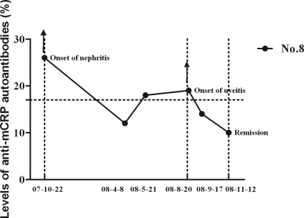
Results: The mCRP autoantibodies were detected in all nine patients with TINU syndrome, significantly higher than that of those with disease controls (P < 0.05). The renal histologic score of mCRP in TINU syndrome was significantly higher than that in disease controls (P < 0.05). The staining of mCRP and human IgG were co-localized in renal and ocular tissues.
Conclusions: It is concluded that mCRP might be a target autoantigen in TINU syndrome.
Figures
References
-
- Mackensen F, Smith JR, Rosenbaum JT: Enhanced recognition, treatment, and prognosis of tubulointerstitial nephritis and uveitis syndrome. Ophthalmology 114: 995–999, 2007 - PubMed
-
- Dobrin RS, Vernier RL, Fish AL: Acute eosinophilic interstitial nephritis and renal failure with bone marrow-lymph node granulomas and anterior uveitis. A new syndrome. Am J Med 59: 325–333, 1975 - PubMed
-
- Mandeville JT, Levinson RD, Holland GN: The tubulointerstitial nephritis and uveitis syndrome. Surv Ophthalmol 46: 195–208, 2001 - PubMed
-
- Gafter U, Kalechman Y, Zevin D, Korzets A, Livni E, Klein T, Sredni B, Levi J: Tubulointerstitial nephritis and uveitis: Association with suppressed cellular immunity. Nephrol Dial Transplant 8: 821–826, 1993 - PubMed
-
- Yoshioka K, Takemura T, Kanasaki M, Akano N, Maki S: Acute interstitial nephritis and uveitis syndrome: Activated immune cell infiltration in the kidney. Pediatr Nephrol 5: 232–234, 1991 - PubMed
Publication types
MeSH terms
Substances
Supplementary concepts
LinkOut - more resources
Full Text Sources
Other Literature Sources
Research Materials
Miscellaneous
