. 2010 Nov 5;285(45):34309-18.
doi: 10.1074/jbc.M110.135822. Epub 2010 Sep 2.

Domain structure of virulence-associated response regulator PhoP of Mycobacterium tuberculosis: role of the linker region in regulator-promoter interaction(s)

Affiliations

Domain structure of virulence-associated response regulator PhoP of Mycobacterium tuberculosis: role of the linker region in regulator-promoter interaction(s)

Anuj Pathak et al. J Biol Chem. .

Abstract

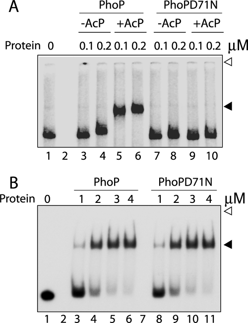
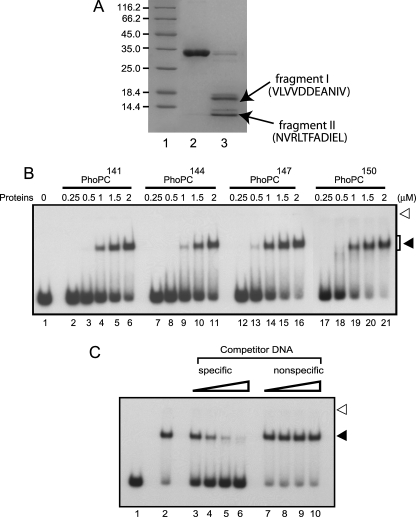
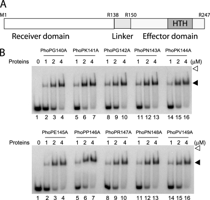
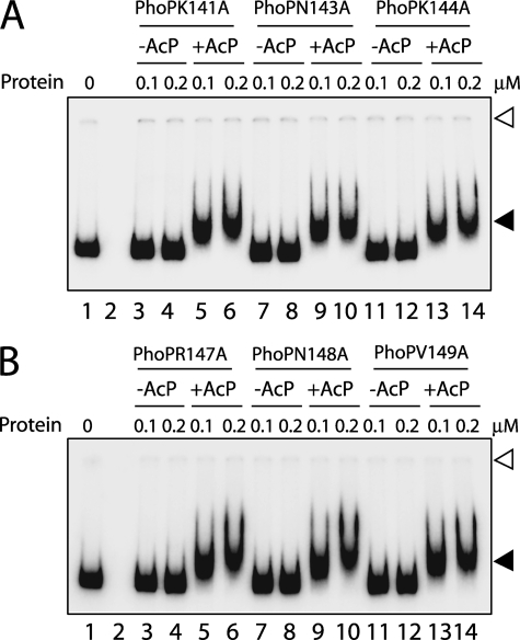
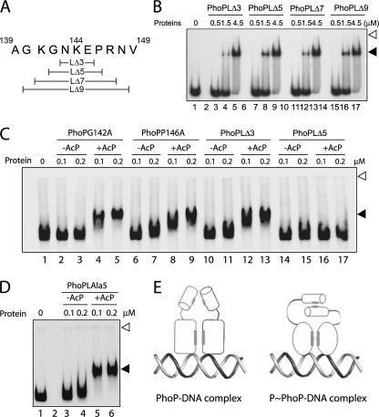
The PhoP and PhoR proteins from Mycobacterium tuberculosis form a highly specific two-component system that controls expression of genes involved in complex lipid biosynthesis and regulation of unknown virulence determinants. The several functions of PhoP are apportioned between a C-terminal effector domain (PhoPC) and an N-terminal receiver domain (PhoPN), phosphorylation of which regulates activation of the effector domain. Here we show that PhoPN, on its own, demonstrates PhoR-dependent phosphorylation. PhoPC, the truncated variant bearing the DNA binding domain, binds in vitro to the target site with affinity similar to that of the full-length protein. To complement the finding that residues spanning Met(1) to Arg(138) of PhoP constitute the minimal functional PhoPN, we identified Arg(150) as the first residue of the distal PhoPC domain capable of DNA binding on its own, thereby identifying an interdomain linker. However, coupling of two functional domains together in a single polypeptide chain is essential for phosphorylation-coupled DNA binding by PhoP. We discuss consequences of tethering of two domains on DNA binding and demonstrate that linker length and not individual residues of the newly identified linker plays a critical role in regulating interdomain interactions. Together, these results have implications for the molecular mechanism of transmission of conformation change associated with phosphorylation of PhoP that results in the altered DNA recognition by the C-terminal domain.


Figures

FIGURE 1.
FIGURE 1.
Phosphorylation-coupled DNA binding by M. tuberculosis PhoP. A, EMSA of radiolabeled promoter region of msl3 for binding of PhoP (lanes 3–6) or PhoPD71N (lanes 7–10) preincubated in phosphorylation mix with or without AcP. B, EMSA of radiolabeled DR1,2 probe with PhoP (lanes 3–6) and PhoPD71N (lanes 8–11). DNA-protein complexes were analyzed by native PAGE and detected by autoradiography as described under “Experimental Procedures.” Note that the bound complexes resolved from the unbound probes differently in A and B because the DNA probes used were of very different size, a 410-bp full-length promoter and a 60-bp oligonucleotide-based duplex DNA in A and B, respectively. Open and filled arrows, origins of the polyacrylamide gel and retarded complexes (with band shifts in the presence of PhoP proteins), respectively. The gels are representative of at least three independent experiments.
FIGURE 2.
FIGURE 2.
Domain structure of PhoP. A, schematic presentation of bipartite domain structure of M. tuberculosis PhoP showing an N-terminal receiver domain (empty), which comprises the site of phosphorylation (Asp-71 for PhoP) (10) and a relatively shorter C-terminal transactivation domain (gray), which includes the structurally characterized helix-turn-helix DNA binding motif (HTH; dark gray; Protein Data Bank entry 2PMU) (11). Truncated PhoP domains were generated by fragmenting PhoP at Lys141, as indicated by an arrow. B, PhoP, PhoPN141, and PhoPC141 (2 μg/lane), expressed and purified as described under “Experimental Procedures,” were analyzed by SDS-polyacrylamide gel electrophoresis and visualized by Coomassie Blue staining. As a reference, molecular mass markers are resolved in lane 1, and the sizes in kDa are indicated to the left. C, purified PhoP (lanes 2–4) and PhoPN141 (lanes 5–7) at ∼3 μm concentration were compared in transphosphorylation assays for their abilities to accept labeled phosphate from ∼3 μm radiolabeled PhoRC in phosphorylation mix at 18 °C for 30, 60, and 120 s, respectively. As a reference, labeled PhoRC was resolved in lane 1. Products were analyzed by denaturing PAGE and detected by autoradiography. D, EMSA of radiolabeled DR1,2 DNA for binding of ∼3.0 μm PhoPC141 in the absence of any competitor (lane 2), in the presence of increasing concentrations of 6.25, 12.5, 25, and 50-fold excess of specific (lanes 3–6) or nonspecific (lanes 7–10) competitor DNA. E, EMSA of radiolabeled promoter region of msl3 for binding of PhoPC141 (lanes 2 and 3). In all cases, protein-DNA complexes were visualized by autoradiography and quantified by scanning the gels on a PhosphorImager. Open and filled arrowheads indicate origins of the polyacrylamide gel and retarded complexes, respectively.
FIGURE 3.
FIGURE 3.
Defining the minimal N-terminal domain of PhoP. A, the indicated N-terminal fragments of PhoP (∼3 μm), expressed and purified as described under “Experimental Procedures,” were compared in phosphotransfer assays with radiolabeled PhoRC for 30, 60, and 120 s, respectively. Reaction conditions, sample analysis, and detection of phosphorylated proteins were as described in the legend to Fig. 2C; labeled PhoRC was resolved in lane 1. B, ClustalW alignment of the amino acid sequences of N-terminal domain of PhoP and its orthologs from E. coli (EcOmpR) and Streptomyces coelicolor (ScPhoP). Conserved and similar amino acids are indicated by black and gray shadings, and α-helices and β-sheets are indicated by filled arrows and empty rectangles, respectively. Six highly conserved residues of the protein subfamily important for phosphorylation and signal propagation are indicated by asterisks.
FIGURE 4.
FIGURE 4.
Defining the minimal C-terminal DNA binding domain of PhoP. A, proteolysis of PhoP was carried out with trypsin, and samples were resolved by SDS-PAGE and visualized by Coomassie Blue staining. Trypsin cleavage sites were identified by N-terminal sequencing of peptides from lane 3 (see “Results” for a description of proteolytic fragments) as described under “Experimental Procedures.” Untreated PhoP was resolved in lane 2; the sizes of marker proteins (lane 1) in kDa are indicated to the left. B, EMSA of radiolabeled DR1,2 DNA for binding of increasing concentrations of indicated PhoPC constructs, expressed and purified as described under “Experimental Procedures.” C, EMSA of radiolabeled DR1,2 DNA for binding of 3.0 μm PhoPC150 (residues 150–247) in the absence of any competitor (lane 2), in the presence of a 6.25-, 12.5-, 25-, and 50-fold molar excess of specific (lanes 3–6) or nonspecific (lanes 7–10) competitor DNA as described in the legends to Fig. 2D. Open and filled arrows indicate origins of the polyacrylamide gel and retarded complexes with band shifts produced in presence of PhoP proteins, respectively.
FIGURE 5.
FIGURE 5.
Alanine-scanning mutagenesis of PhoP linker region. A, revised domain structure of M. tuberculosis PhoP showing newly identified linker region (residues 139–149) shown in light gray. Note that Arg138 and Arg150 represent the last residue of the proximal N-terminal domain and the first residue of the distal C-terminal domain of PhoP, respectively. B, EMSA of radiolabeled DR1,2 DNA for binding of the indicated single alanine mutants of PhoP, expressed and purified as described under “Experimental Procedures.” Sample analysis and detection of protein-DNA complexes were as described in the legend to Fig. 1B.
FIGURE 6.
FIGURE 6.
A and B, EMSA of radiolabeled msl3 promoter region for binding of the indicated PhoP linker mutants carrying single alanine substitutions. Sample analysis and detection of protein-DNA complexes were as described in the legend to Fig. 1. Open and filled arrows indicate origins of the polyacrylamide gel and retarded complexes with band shifts produced in the presence of PhoP proteins, respectively. The gels are representative of at least three independent experiments.
FIGURE 7.
FIGURE 7.
A, scheme showing the linker deletion constructs of M. tuberculosis PhoP. B, EMSA of radiolabeled DR1,2 DNA for binding of PhoP linker deletion mutants. Sample analysis and detection of protein-DNA complexes were as described in the legend to Fig. 1A. C, EMSA of radiolabeled msl3 promoter region for binding of the indicated single alanine mutants as well as linker deletion mutants of PhoP, preincubated in phosphorylation mix with or without AcP. D, EMSA of radiolabeled msl3 promoter region for binding of PhoPLAla5 mutant, preincubated in phosphorylation mix with or without AcP. Each of the gels is representative of at least three independent experiments with at least two different preparations of protein stocks. Sample analysis and detection of protein-DNA complexes were as described in the legends to Fig. 1A. E, dual mode of DNA binding by unphosphorylated and phosphorylated PhoP, respectively, employing mechanisms that are either linker-independent or require the presence of at least eight residues of the PhoP linker (see “Results”). The N-terminal domain is represented as a cylinder, and the C-terminal DNA binding domain is represented as an ellipse for PhoP and a circle for phosphorylated PhoP (to suggest phosphorylation-coupled conformational change), respectively; the protein-protein interaction interface is shaded in gray.

Similar articles

Cited by

References

    1. Fontan P. A., Walters S., Smith I. (2004) Curr. Sci. 86, 122–134
    1. Av-Gay Y., Deretic V. (2005) in Tuberculosis and the Tubercle Bacillus (Cole S. T. ed) pp. 359–367, American Society for Microbiology Press, Washington, D. C.
    1. Pérez E., Samper S., Bordas Y., Guilhot C., Gicquel B., Martín C. (2001) Mol. Microbiol. 41, 179–187 - PubMed
    1. Walters S. B., Dubnau E., Kolesnikova I., Laval F., Daffe M., Smith I. (2006) Mol. Microbiol. 60, 312–330 - PubMed
    1. Gonzalo Asensio J., Maia C., Ferrer N. L., Barilone N., Laval F., Soto C. Y., Winter N., Daffé M., Gicquel B., Martín C., Jackson M. (2006) J. Biol. Chem. 281, 1313–1316 - PubMed

Publication types

MeSH terms

LinkOut - more resources