Case Reports
doi: 10.4103/0974-2069.64364.
Ductal aneurysm masquerading as nonresolving pneumonia: A challenging differential!
Affiliations
- PMID: 20814482
- PMCID: PMC2921525
- DOI: 10.4103/0974-2069.64364
Item in Clipboard
Case Reports
Ductal aneurysm masquerading as nonresolving pneumonia: A challenging differential!
Ann Pediatr Cardiol.
2010 Jan.
Abstract
We report here, the case of a six-and-a-half-month-old boy investigated for persistent respiratory distress and homogeneous opacity in the left upper lobe. Echocardiography revealed a giant ductal aneurysm compressing the left pulmonary artery and upper lobe division of the left bronchus. Computerized tomography angiogram delineated the exact anatomy and prompt surgical resection provided a successful cure to this lesser known entity.
Keywords: Ductal aneurysm; PDA; pneumonia.
Conflict of interest statement
Figures
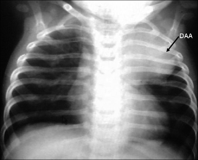
Chest X-ray (PA view) showing homogenous rounded opacity in the left upper thorax produced by the ductal aneurysm, normal lung parenchyma, normal pulmonary blood flow, and there was no cardiomegaly. DAA-ductus arteriosus aneurysm

Two-dimensional echocardiography from modified suprasternal echocardiographic long axis view showing the aneurysm and the ampulla (arrow)

Two-dimensional echocardiography from modified suprasternal short axis view showing large ductal aneurysm with wide ampulla

High parasternal short axis view with color flow mapping showing tiny ductal flow (left to right) with normal flow in the right pulmonary artery DAA-ductus arteriosus aneurysm, RPA-right pulmonary artery, LPA-left pulmonary artery, DAo-descening aorta. Ao arch-aortic arch, PDA-patent ductus arteriosus
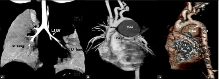
High resolution computed tomography (HRCT) angiography of the chest (a) Compression of left upper lobe bronchus and left upper lobe collapse (b) Large ductal aneurysm compressing the left pulmonary artery (c) Three-dimensional reconstruction of HRCT chest from the posterior aspect, showing the large ductal aneurysm originating from the descending aorta just distal to the origin of the left subclavian artery. Lt Br-left bronchus, rt lung-right lung, lt lung-left lung, AAo-ascending aorta, Dao-descending aorta, LPA-left pulmonary artery, DAA-ductus arteriosus aneurysm
References
-
- Lund JT, Jensen MB, Hjelms E. Aneurysm of the ductus arteriosus.A review of the literature and the surgical implications. Eur J Cardiothorac Surg. 1991;5:566–70. - PubMed
-
- Varma PK, Vallath G, Neema PK, Sinha PK, Sivadasanpillai H, Menon MU, et al. Clinical profile of post-operative ductal aneurysm and usefulness of sternotomy and circulatory arrest for its repair. Eur J Cardiothorac Surg. 2005;27:416–9. - PubMed
-
- Zilinskas VJ, Maknavicius S, Baliulis G. Successful resection of ductus arteriosus aneurysm in infancy. Ann Thorac Surg. 2000;69:282–3. - PubMed
-
- Stewart A, Dyamenahalli U, Greenberg SB, Drummond-Webb J. Ductus arteriosus aneurysm with community-acquired methicillin-resistant Staphylococcus aureus infection and spontaneous rupture: A potentially fatal quandary. Pediatrics. 2006;117:e1259–62. - PubMed
-
- Dyamenahalli U, Smallhorn JF, Geva T, Fouron JC, Cairns P, Jutras L, et al. Isolated ductus arteriosus aneurysm in the fetus and infant: A multi-institutional experience. J Am Coll Cardiol. 2000;36:262–9. - PubMed