

Case Reports
doi: 10.1155/2010/175613.
Epub 2010 Aug 12.
Pathology of macular foveoschisis associated with degenerative myopia
Affiliations
- PMID: 20814547
- PMCID: PMC2931386
- DOI: 10.1155/2010/175613
Item in Clipboard
Case Reports
Pathology of macular foveoschisis associated with degenerative myopia
J Ophthalmol.
2010.
Abstract
This is a clinicopathological paper on the histologic findings in myopia-associated macular foveoschisis. The findings on ophthalmic pathological study of a 73-year-old woman with high myopia are reviewed. Multiple retinoschisis cavities involving both the macula and retinal periphery were disclosed. Our paper offers tissue evidence and supports recent ocular coherence tomography reports of eyes with high myopia and associated macular foveoschisis.
Figures
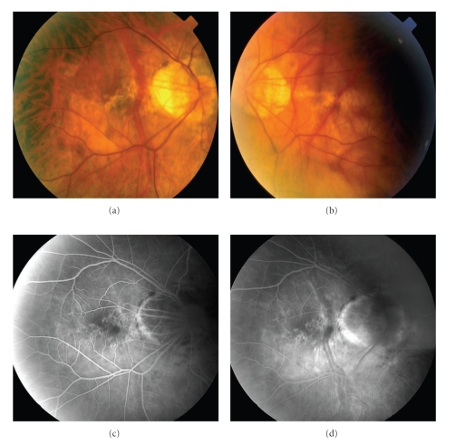
(a) Clinical photographs of the fundus of the right and (b) left eyes. Bilateral optic nerve crescents and staphylomas are seen in both eyes. There are also degenerative changes seen in the macular areas of both eyes. (c) Fluorescein angiogram of the right eye in the early (a) and late (b) phases demonstrating staining of the optic nerve crescents and small window defects along the macular region.
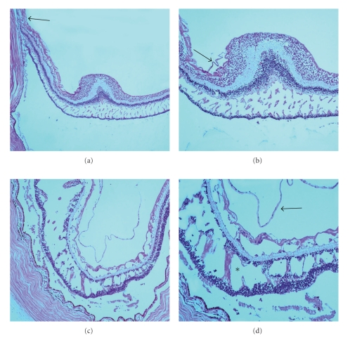
(a) Photomicrograph of the right eye demonstrating areas of macular foveoschisis. A region containing the staphyloma is also seen (black arrow). (b) Higher magnification of macular foveoschisis seen in multiple layers of the retina including the outer plexiform layer, inner plexiform layer, nerve fiber layer, and the outer plexiform layer in the perifoveal region. A thin fibrous preretinal membrane is seen (black arrow). (hematoxylin and eosin, original magnification, (a) x50; (b) x100). (c) Photomicrograph of the left eye demonstrating classical retinoschisis in the outer plexiform layer, ganglion cell layer, and nerve fiber layer. (d) Higher magnification demonstrating neuronal bridges between both nuclear layers (asterisk). A fibrous preretinal membrane is seen (black arrow). (hematoxylin and eosin, original magnification, (a) x50; (b) x100).
Similar articles
-
Relationship between macular bending and foveoschisis in myopic patients.Optom Vis Sci. 2014 May;91(5):497-506. doi: 10.1097/OPX.0000000000000250. Optom Vis Sci. 2014. PMID: 24727824
-
Structural profile of dome-shaped macula in degenerative myopia and its association with macular disorders.BMC Ophthalmol. 2020 May 24;20(1):202. doi: 10.1186/s12886-020-01473-2. BMC Ophthalmol. 2020. PMID: 32448138 Free PMC article.
-
[Factors linked to foveoschisis in high myopia].J Fr Ophtalmol. 2014 Feb;37(2):138-42. doi: 10.1016/j.jfo.2013.11.001. Epub 2014 Jan 31. J Fr Ophtalmol. 2014. PMID: 24486073 French.
-
Myopic foveoschisis: a clinical review.Eye (Lond). 2015 May;29(5):593-601. doi: 10.1038/eye.2014.311. Epub 2015 Mar 6. Eye (Lond). 2015. PMID: 25744445 Free PMC article. Review.
-
Macular buckle technique in myopic traction maculopathy: a 16-year review of the literature and a comparison with vitreous surgery.Graefes Arch Clin Exp Ophthalmol. 2018 May;256(5):863-877. doi: 10.1007/s00417-018-3947-3. Epub 2018 Mar 28. Graefes Arch Clin Exp Ophthalmol. 2018. PMID: 29589106 Review.
Cited by
-
Peripapillary retinoschisis in glaucomatous eyes.PLoS One. 2014 Feb 28;9(2):e90129. doi: 10.1371/journal.pone.0090129. eCollection 2014. PLoS One. 2014. PMID: 24587238 Free PMC article.
-
Different foveal schisis patterns in each retinal layer in eyes with hereditary juvenile retinoschisis evaluated by en-face optical coherence tomography.Graefes Arch Clin Exp Ophthalmol. 2017 Apr;255(4):719-723. doi: 10.1007/s00417-016-3552-2. Epub 2016 Nov 16. Graefes Arch Clin Exp Ophthalmol. 2017. PMID: 27853955
-
Factors Determining the Morphology of Peripapillary Retinoschisis.Clin Ophthalmol. 2021 Mar 25;15:1293-1300. doi: 10.2147/OPTH.S301196. eCollection 2021. Clin Ophthalmol. 2021. PMID: 33790537 Free PMC article.
-
Peripapillary Retinoschisis in Glaucoma Patients.J Ophthalmol. 2016;2016:1612720. doi: 10.1155/2016/1612720. Epub 2016 Mar 16. J Ophthalmol. 2016. PMID: 27069674 Free PMC article.
-
IMI Pathologic Myopia.Invest Ophthalmol Vis Sci. 2021 Apr 28;62(5):5. doi: 10.1167/iovs.62.5.5. Invest Ophthalmol Vis Sci. 2021. PMID: 33909033 Free PMC article. Review.
References
-
- Takano M, Kishi S. Foveal retinoschisis and retinal detachment in severely myopic eyes with posterior staphyloma. American Journal of Ophthalmology. 1999;128(4):472–476. - PubMed
-
- Akiba J, Konno S, Sato E, Yoshida A. Retinal detachment and retinoschisis detected by optical coherence tomography in myopic eye with a macular hole. Ophthalmic Surgery and Lasers. 2000;31(3):240–242. - PubMed
-
- Ikuno Y, Gomi F, Tano Y. Potent retinal arteriolar traction as a possible cause of myopic foveoschisis. American Journal of Ophthalmology. 2005;139(3):462–467. - PubMed
-
- Baba T, Ohno-Matsui K, Futagami S, et al. Prevalence and characteristics of foveal retinal detachment without macular hole in high myopia. American Journal of Ophthalmology. 2003;135(3):338–342. - PubMed
Publication types
LinkOut - more resources
Full Text Sources
