. 2010 Aug 30;5(8):e12479.
doi: 10.1371/journal.pone.0012479.

TNF inhibits Notch-1 in skeletal muscle cells by Ezh2 and DNA methylation mediated repression: implications in duchenne muscular dystrophy

Affiliations

TNF inhibits Notch-1 in skeletal muscle cells by Ezh2 and DNA methylation mediated repression: implications in duchenne muscular dystrophy

Swarnali Acharyya et al. PLoS One. .

Abstract

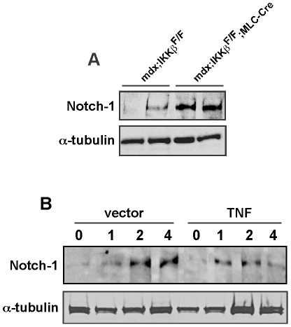
Background: Classical NF-kappaB signaling functions as a negative regulator of skeletal myogenesis through potentially multiple mechanisms. The inhibitory actions of TNFalpha on skeletal muscle differentiation are mediated in part through sustained NF-kappaB activity. In dystrophic muscles, NF-kappaB activity is compartmentalized to myofibers to inhibit regeneration by limiting the number of myogenic progenitor cells. This regulation coincides with elevated levels of muscle derived TNFalpha that is also under IKKbeta and NF-kappaB control.

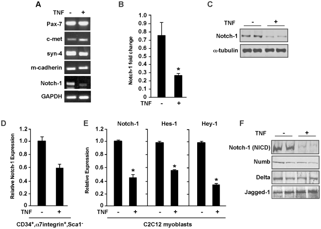
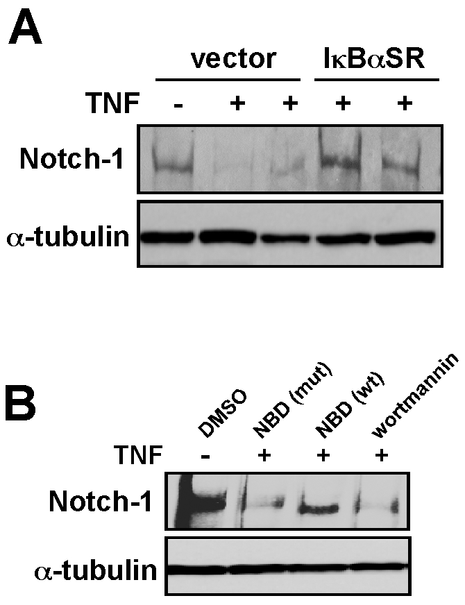
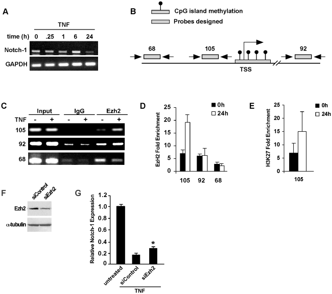
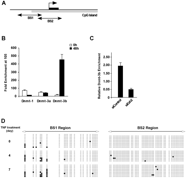
Methodology/principal findings: Based on these findings we speculated that in DMD, TNFalpha secreted from myotubes inhibits regeneration by directly acting on satellite cells. Analysis of several satellite cell regulators revealed that TNFalpha is capable of inhibiting Notch-1 in satellite cells and C2C12 myoblasts, which was also found to be dependent on NF-kappaB. Notch-1 inhibition occurred at the mRNA level suggesting a transcriptional repression mechanism. Unlike its classical mode of action, TNFalpha stimulated the recruitment of Ezh2 and Dnmt-3b to coordinate histone and DNA methylation, respectively. Dnmt-3b recruitment was dependent on Ezh2.

Conclusions/significance: We propose that in dystrophic muscles, elevated levels of TNFalpha and NF-kappaB inhibit the regenerative potential of satellite cells via epigenetic silencing of the Notch-1 gene.


Conflict of interest statement

Competing Interests: The authors have declared that no competing interests exist.

Figures

Figure 1
Figure 1. TNF regulates Notch-1 expression.
A. Mononuclear cell cultures were prepared from mdx muscles and cultured under proliferation conditions and either left untreated or treated with TNF. After 24 hrs cells were harvested and processed for total RNA, and subsequently semi-quantitative RT-PCR reactions were performed probing for satellite cell markers. GAPDH was used as a control. B. Quantitative real-time RT-PCR analysis of Notch-1 from satellite cell cultures untreated and treated with TNF. Values shown were normalized to GAPDH levels. Asterisk denotes statistical significance, p = 0.03708. C. Western blot analysis probing for total Notch-1 receptor from duplicate mdx satellite cell cultures treated with TNF. The blot was stripped and re-probed for α-tubulin used as a loading control D. Quantitative real-time RT-PCR was repeated as in (B) from FACS sorted mononuclear cells enriched for a CD34+, α7integrin+, Sca1 population. E. C2C12 myoblasts were cultured under proliferating conditions and either not treated or treated with TNF. Total RNA was prepared and quantitative RT-PCR analysis was performed probing for Notch-1 and Notch-1 targets, Hes-1 and Hey-1. Asterisk denotes statistical significance; Notch-1, p = 0.04844; Hes-1, p = 0.001; Hey-1, p = 0.0149. F. C2C12 myoblasts were cultured in treated in duplicate with TNF and protein lysates were subsequently probed with the intercellular activated form of Notch-1 (NICD), or with Notch signaling components, Delta and Jagged-1.
Figure 2
Figure 2. TNF regulates Notch-1 expression in vivo.
A. Muscle homogenates were prepared from 7–8 week old mdx mice that either contained or lacked IKKβ and western analysis was performed probing for Notch-1. The blot was stripped and reprobed for α-tubulin used as a loading control. B. Nude mice were implanted with vector control CHO cells or CHO cells expressing TNF. Once tumors were established, muscle injury was induced to tibialis anterior muscles with cardiotoxin injections. At indicated days post-toxin injections, muscles were harvested and homogenates prepared for western analysis probing for Notch-1 and α-tubulin as a loading control.
Figure 3
Figure 3. TNF regulation of Notch-1 is dependent on NF-κB.
A. Western analysis of Notch-1 was performed in either untreated or TNF treated C2C12 vector myoblasts or myoblasts stably expressing the IκBα-SR transgene. B. C2C12 myoblasts were pre-treated for 1 hr with either DMSO, an IKK inhibitor peptide (NBD) in both a wild type (wt) and inactive mutant form (mut), or with a treatment of wortmannin. Following TNF treatment, cells were harvested and western was performed probing for Notch-1 and α-tubulin as a control.
Figure 4
Figure 4. TNF repression of Notch-1 occurs through the recruitment of Ezh2.
A. Time course analysis of Notch-1 expression following TNF treatment of C2C12 myoblasts. B. Schematic illustration of the Notch-1 gene indicating conserved regions of CpG content (blue bars) that are located in positions proximal to the transcriptional start site (TSS), and where probes were designed for ChIP analysis. Also indicated is a CpG island immediately surrounding the TSS (red bar). C, D. C2C12 myoblasts were treated with TNF and at indicated times cells were harvested and subsequently prepared for ChIP analysis probing for Ezh2 by both semi-quantitative (C) and quantitative (D) RT-PCR. E. Using cell extracts prepared in C, additional ChIPs were performed for methylation of H3K27. F. C2C12 myoblasts were transfected with scrambled siRNA (siControl) or siRNA targeting Ezh2 (siEzh2) and following 48 hrs, western blot analyze was performed probing for Ezh2 and α-tubulin used as a loading control. G. C2C12 myoblasts transfections were performed with siControl and siEzh2 oligonucleotides and next day cells were treated for TNF for 48 hrs, and subsequently processed for Notch-1 expression by real-time RT-PCR. Asterisks denotes p = 0.00314).
Figure 5
Figure 5. TNF promotes Dnmt-3b recruitment and DNA methylation on the Notch-1 promoter.
A. Schematic illustration of the Notch-1 promoter indicating the CpG islands in the BS1 and BS2 regions proximal to the TSS. B. C2C12 myoblasts were treated with TNF for 48 hrs at which time cells were prepared for quantitative ChIP analysis for Dnmt-1, Dnmt-3a, and Dnmt-3b. C. C2C12 myoblasts were transfected with scrambled siRNA or siRNA against Ezh2 and the following day cells were treated for TNF for 48 hrs and ChIP analysis for Dnmt-3b was subsequently performed. D. C2C12 myoblasts were treated with TNF for up to 7 days and at indicated times, cells were processed for bisulfite sequencing of the BS1 region within the Notch-1 promoter. Note the increase in BS1 methylation at discrete CpG dinucleotides over time with TNF treatment.

References

    1. Le Grand F, Rudnicki M. Satellite and stem cells in muscle growth and repair. Development. 2007;134:3953–3957. - PubMed
    1. Peault B, Rudnicki M, Torrente Y, Cossu G, Tremblay JP, et al. Stem and progenitor cells in skeletal muscle development, maintenance, and therapy. Mol Ther. 2007;15:867–877. - PubMed
    1. Davies KE, Nowak KJ. Molecular mechanisms of muscular dystrophies: old and new players. Nat Rev Mol Cell Biol. 2006;7:762–773. - PubMed
    1. Kuang S, Rudnicki MA. The emerging biology of satellite cells and their therapeutic potential. Trends Mol Med. 2008;14(2):82–91. - PubMed
    1. Khurana TS, Davies KE. Pharmacological strategies for muscular dystrophy. Nat Rev Drug Discov. 2003;2:379–390. - PubMed

Publication types

MeSH terms

LinkOut - more resources