. 2010 Nov;67(11):715-28.
doi: 10.1002/cm.20482.

Coupling between microtubule sliding, plus-end growth and spindle length revealed by kinesin-8 depletion

Affiliations

Coupling between microtubule sliding, plus-end growth and spindle length revealed by kinesin-8 depletion

Haifeng Wang et al. Cytoskeleton (Hoboken). 2010 Nov.

Abstract

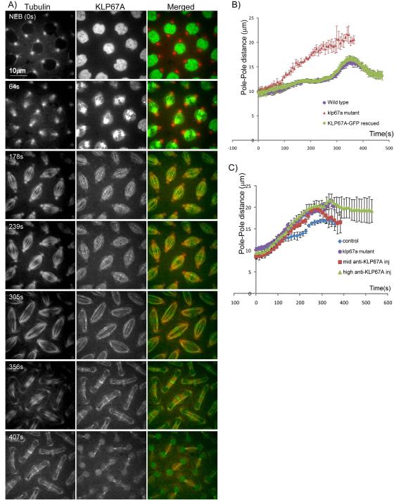
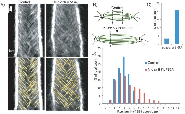
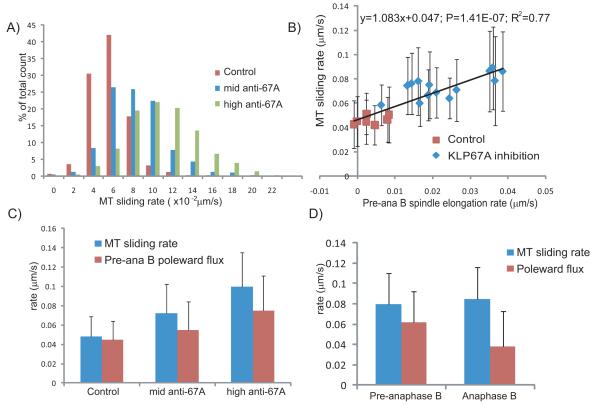
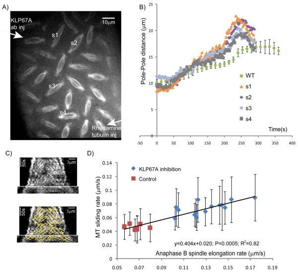
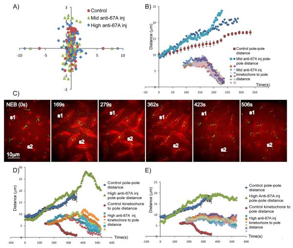
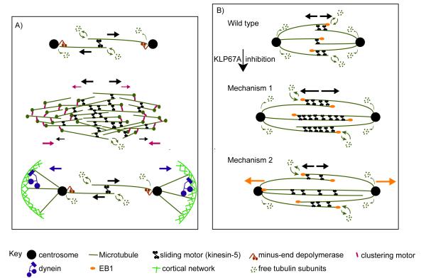
Mitotic spindle length control requires coordination between microtubule (MT) dynamics and motor-generated forces. To investigate how MT plus-end polymerization contributes to spindle length in Drosophila embryos, we studied the dynamics of the MT plus-end depolymerase, kinesin-8, and the effects of kinesin-8 inhibition using mutants and antibody microinjection. As expected, kinesin-8 was found to contribute to anaphase A. Furthermore, kinesin-8 depletion caused: (i) excessive polymerization of interpolar (ip) MT plus ends, which "overgrow" to penetrate distal half spindles; (ii) an increase in the poleward ipMT sliding rate that is coupled to MT plus-end polymerization; (iii) premature spindle elongation during metaphase/anaphase A; and (iv) an increase in the anaphase B spindle elongation rate which correlates linearly with the MT sliding rate. This is best explained by a revised "ipMT sliding/minus-end depolymerization" model for spindle length control which incorporates a coupling between ipMT plus end dynamics and the outward ipMT sliding that drives poleward flux and spindle elongation.


Figures

Figure 1
Figure 1. Localization and dynamics of KLP67A-GFP in Drosophila embryo mitotic spindles
(A) Micrographs from a time lapse video of a representative embryo showing rhodamine-tubulin (left) KLP67A-GFP (center) and merged (right) (tubulin:red; KLP67A-GFP:green). Time in each frame is given in seconds from nuclear envelope breakdown (NEB). In interphase and prophase, KLP67A-GFP was sequestered in the nucleus. Starting after NEB through telophase, KLP67A-GFP localized all along spindle MTs and faintly decorated astral MTs. During metaphase and anaphase, we also observed distinct spots of KLP67A-GFP that lay near the metaphase spindle equator and separated with the chromosomes during anaphase A. This is likely to be kinetochore-associated KLP67A as reported previously by others (Savoian and Glover, 2010). During anaphase B and telophase, KLP67A-GFP was concentrated at the spindle midzone. Once the nuclear envelope reformed, KLP67A-GFP was re-recruited into the nucleus. (B) Spindle pole dynamics in (i) wild type embryos; (ii) klp67A mutant embryos; and (iii) KLP67A-GFP-rescued klp67A mutant embryos showing that the expression of KLP67A-GFP fully compensated for the depletion of KLP67A in the mutants. (C) Spindle pole dynamics in wild type embryos; klp67A mutant embryos; the “mid anti-KLP67A”- injected embryos; and the “high anti-KLP67A” injected embryos showing that KLP67A depletion by anti-KLP67A microinjection produced premature spindle pole separation, virtually identical to the klp67A hypomorphic mutants. See Fig. S1 for still images.
Figure 2
Figure 2. KLP67A depletion affects the dynamics of growing MT plus-ends
(A) Comparison of representative EB1-GFP kymographs from control and “mid anti-KLP67A” injected embryos showing that KLP67A depletion allows more growing MTs to cross the spindle equator and enter the distal half spindle to reach the opposite pole. (B) Cartoon showing change in spindle MT organization after KLP67A inhibition. MTs are shown in green and centrosomes are shown in black. Dotted black line shows the arbitrary division of the spindle into three domains along the pole-pole axis which were used to semi-quantify the extent of penetration of one half spindle by MT plus-ends from the other half spindle. (C) Comparison of the percentage of growing MT plus ends that penetrate into the distal one-third spindle region, adjacent to the distal pole (division into the three regions as shown in B) - control and “mid” anti-KLP67A injected embryos are shown. (D) Histogram of EB1 speckle run lengths in control and “mid” anti-KLP67A injected embryos. Average length and total counts for C and D are shown in Table 1.
Figure 3
Figure 3. KLP67A depletion increased the rates of (i) ipMT sliding away from the spindle equator; and (ii) poleward MT flux
(A) Histogram of MT sliding rate (tubulin speckle velocity away from the equator) in control (red), the “mid” anti-KLP67A injected embryos (blue) and the “high” anti-KLP67A injected embryos (green). Total counts are shown in Table 1. (B) Linear correlation between the MT sliding rate and the premature pre-anaphase B spindle elongation rate in each spindle from control and anti-KLP67A injected embryos. P-value =1.41499E-07. R squared = 0.77. For each spindle, 50-200 speckles were measured. (C) Comparison of the MT sliding rate (blue) and pre-anaphase B MT poleward flux rate (red) in controls compared to “mid” and “high” anti-KLP67A injected embryos. P<2.2e-16 for all pairwise t-tests. Total counts are shown in Table 1. (D) Comparison of MT sliding rate (blue) and MT poleward flux rate (red) during pre-anaphase B (911 speckles in 8 spindles measured) and anaphase B (877 speckles in the same 8 spindles measured) in a representative embryo injected with “mid” anti-KLP67A antibody.
Figure 4
Figure 4. Higher anaphase B spindle elongation rate after KLP67A depletion is correlated with an increase in the MT sliding rate
(A) A still image from a time lapse video of a representative embryo injected with anti-KLP67A antibody (at the upper left corner) showing a gradient of mitotic defects. (B) Spindle pole dynamics of the four spindles shown in A compared to controls (WT) showing that KLP67A inhibition caused an increase in anaphase B spindle elongation rate. (C). Representative tubulin kymograph from an embryo partially depleted of KLP67A, showing that tubulin speckles move away from the spindle equator at a similar rate to the rate of spindle pole separation during anaphase B. (D) Linear correlation between the MT sliding rate and anaphase B spindle elongation rate in spindles. Data from two representative embryos displaying a higher anaphase B spindle elongation rate after partial KLP67A depletion compared to a control embryo. P-value=0.0005.R square=0.82. For each spindle, 50-200 speckles were measured.
Figure 5
Figure 5. Chromosome congression and anaphase A are perturbed by anti-KLP67A antibody injection
(A) Positions of metaphase kinetochores in: (i) control (red); (ii) “mid” anti-KLP67A (green); and (iii) “high” anti-KLP67A (blue) injected embryo spindles, showing that KLP67A inhibition causes kinetochores to scatter over a wider range relative to the spindle equator. Average pole positions are plotted along x-axis. (B) Pole-pole distance and kinetochore-to-pole distances as a function of time for two representative spindles injected with “mid” concentrations of anti-KLP67A in comparison with controls (red). After “mid” anti-KLP67A injection, anaphase A started at the normal time, kinetochores moved synchronously toward the pole, and chromosome segregation was completed. Based on their different behavior in anaphase B spindle elongation, spindles injected with mid concentration of anti-KLP67A could be categorized into two groups. In the first group (resembled by pole dynamics plotted in blue), the onset of anaphase B is blurred because there’s no significant difference between pre-anaphase B and anaphase B spindle elongation rate. In the second group (resembled by the pole dynamics plotted in light blue), the anaphase B spindle elongation rate is usually higher than control, the timing of anaphase A and anaphase B is perturbed. (C, D ,E) show that “high” anti-KLP67A injection caused slow or incomplete kinetochore to pole movement (C, MT: red; CID-GFP: green), which could be categorized into two groups. In the first group (exemplified by spindle S1 in C and the data plotted in D), kinetochores moved toward the poles in a non-synchronous manner at a slower rate than normal but were nonetheless able to reach the poles to complete their separation. In the second group (exemplified by spindle S2 in C and the data plotted in E), kinetochore-to-pole movement stopped prematurely after an initial phase of separation. Noticeably, in both groups, anaphase A started later than in control spindles.
Figure 6
Figure 6. Models for spindle length control
(A) Diagram of current models for spindle length control. Forces generated by different motors are shown by arrows with corresponding colors. According to the “ipMT sliding and minus-end depolymerization” model (top panel) (Brust-Mascher et al., 2004; Brust-Mascher and Scholey, 2002; Goshima et al., 2005), ipMT sliding driven by MT sliding motors (e.g. kinesin-5) is balanced by MT minus-end depolymerization at the poles to keep the metaphase spindle length constant. At anaphase B onset, downregulation of MT minus-end depolymerization allows ipMT sliding to drive spindle elongation. According to the “slide and cluster” model (center panel) (Burbank et al., 2007; Dumont and Mitchison, 2009), while MT sliding and clustering motors cooperate to drive outward MT movement near the equator (where MT overlaps are mostly anti-parallel), the clustering motors counteract the outwardly sliding MTs near the poles (where MT overlaps are mostly parallel), so the rate of MT sliding gradually slows down with increasing distance from the equator, and the spindle pole is formed when the MT sliding velocity slows to zero. This model focuses on the assembly and maintenance of anastral metaphase spindles rather than anaphase B spindle elongation. The astral MT pulling model (bottom panel) is proposed as a mechanism for spindle elongation (Saunders et al., 2007; Sharp et al., 2000a). According to this model, the cortical forces exerted on astral MTs by dynein generate pulling forces for pole-pole separation and spindle elongation. (B) Possible mechanisms by which excessive ipMT polymerization leads to faster ipMT sliding after KLP67A inhibition. Compared to control spindles (top panel), KLP67A inhibition caused prolonged and excessive ipMT plus end polymerization. In the first mechanism (center panel), we assume that the engaged force-generating sliding motors, regulated by the small and rapidly turning over antiparallel overlaps, are loaded. The abnormally large antiparallel MT overlap zone allows the recruitment of more MT sliding motors, which reduces the load per motor, thereby increasing their sliding velocity. In the second mechanism (bottom panel), we assume that the MT sliding motors work at their unloaded velocity. KLP67A depletion allows “overgrowing” ipMT polymer ratchets to push the distal centrosome outwards (orange arrows). Presumably the MT sliding motors may then serve as brakes that control the faster, MT polymer ratchet dependent ipMT sliding.

Similar articles

Cited by

References

    1. Brust-Mascher I, Civelekoglu-Scholey G, Kwon M, Mogilner A, Scholey JM. Model for anaphase B: role of three mitotic motors in a switch from poleward flux to spindle elongation. Proc Natl Acad Sci U S A. 2004;101:15938–15943. - PMC - PubMed
    1. Brust-Mascher I, Scholey JM. Microtubule flux and sliding in mitotic spindles of Drosophila embryos. Mol Biol Cell. 2002;13:3967–3975. - PMC - PubMed
    1. Brust-Mascher I, Scholey JM. Microinjection Techniques for Studying Mitosis in the Drosophila melanogaster Syncytial Embryo. JoVE. 2009;31 http://www.jove.com/index/details.stp?id=1382, DOI: 10.3791/1382. - PMC - PubMed
    1. Brust-Mascher I, Sommi P, Cheerambathur DK, Scholey JM. Kinesin-5-dependent poleward flux and spindle length control in Drosophila embryo mitosis. Mol Biol Cell. 2009;20:1749–1762. - PMC - PubMed
    1. Burbank KS, Mitchison TJ, Fisher DS. Slide-and-cluster models for spindle assembly. Curr Biol. 2007;17:1373–1383. - PubMed

Publication types

MeSH terms

LinkOut - more resources