

Regional and hemispheric determinants of language laterality: implications for preoperative fMRI
- PMID: 20814960
- PMCID: PMC3193373
- DOI: 10.1002/hbm.21130
Regional and hemispheric determinants of language laterality: implications for preoperative fMRI
Abstract
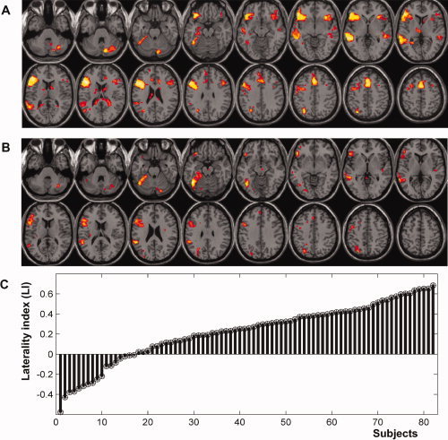
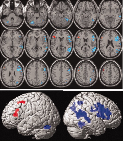
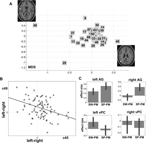
Language is typically a function of the left hemisphere but the right hemisphere is also essential in some healthy individuals and patients. This inter-subject variability necessitates the localization of language function, at the individual level, prior to neurosurgical intervention. Such assessments are typically made by comparing left and right hemisphere language function to determine "language lateralization" using clinical tests or fMRI. Here, we show that language function needs to be assessed at the region and hemisphere specific level, because laterality measures can be misleading. Using fMRI data from 82 healthy participants, we investigated the degree to which activation for a semantic word matching task was lateralized in 50 different brain regions and across the entire cortex. This revealed two novel findings. First, the degree to which language is lateralized across brain regions and between subjects was primarily driven by differences in right hemisphere activation rather than differences in left hemisphere activation. Second, we found that healthy subjects who have relatively high left lateralization in the angular gyrus also have relatively low left lateralization in the ventral precentral gyrus. These findings illustrate spatial heterogeneity in language lateralization that is lost when global laterality measures are considered. It is likely that the complex spatial variability we observed in healthy controls is more exaggerated in patients with brain damage. We therefore highlight the importance of investigating within hemisphere regional variations in fMRI activation, prior to neuro-surgical intervention, to determine how each hemisphere and each region contributes to language processing.
Copyright © 2010 Wiley-Liss, Inc.
Figures
References
-
- Abou‐Khalil B ( 2007): Methods for determination of language dominance: The Wada test and proposed noninvasive alternatives. Curr Neurol Neurosci Rep 7: 483–490. - PubMed
-
- Arora J, Pugh K, Westerveld M, Spencer S, Spencer DD, Todd Constable R ( 2009): Language lateralization in epilepsy patients: fMRI validated with the Wada procedure. Epilepsia 50: 2225–2241. - PubMed
-
- Ashburner J, Friston KJ ( 2005): Unified segmentation. Neuroimage 26: 839–851. - PubMed
-
- Baciu MV, Watson JM, McDermott KB, Wetzel RD, Attarian H, Moran CJ, Ojemann JG ( 2003): Functional MRI reveals an interhemispheric dissociation of frontal and temporal language regions in a patient with focal epilepsy. Epilepsy Behav 4: 776–780. - PubMed
Publication types
MeSH terms
Substances
Grants and funding
LinkOut - more resources
Full Text Sources
Other Literature Sources
