Comparative Study
. 2010 Sep 3:10:477.
doi: 10.1186/1471-2407-10-477.

Expression of CIAPIN1 in human colorectal cancer and its correlation with prognosis

Affiliations
Comparative Study

Expression of CIAPIN1 in human colorectal cancer and its correlation with prognosis

Hai Shi et al. BMC Cancer. .

Abstract

Background: The cytokine-induced anti-apoptotic molecule (CIAPIN1) had been found to be a differentially-expressed gene involved in a variety of cancers, and it was also considered as a candidate tumour suppressor gene in gastric cancer, renal cancer and liver cancer. However, studies on the role of CIAPIN1 in colorectal cancer were still unavailable. The aim of this study was to determine the prognostic impact of CIAPIN1 in 273 colorectal cancer (CRC) samples and to investigate the CIAPIN1 expression in CRC cell lines after inducing differentiation.

Methods: Immunohistochemical analysis was performed to detect the expression of CIAPIN1 in CRC samples from 273 patients. The relationship between CIAPIN1 expression and patients' characteristics (gender, age, location of cancer, UICC stage, local recurrence and tumour grade factors) was evaluated. In addition, these patients were followed up for five consecutive years to investigate the relationship between CIAPIN1 expression and the prognosis of CRC. We induced the differentiation of the CRC cell lines HT29 and SW480, in order to detect the expression of CIAPIN1 in the process of CRC cells differentiation.

Results: Results indicated that CIAPIN1 was mainly expressed in the cytoplasm and nucleus, and that its expression level in cancer samples was significantly lower than in normal tissues. The Wilcoxon-Mann-Whitney test showed a significant difference in the differential expression of CIAPIN1 in patients with different T and UICC stages, and tumour grade (P = 0.0393, 0.0297 and 0.0397, respectively). The Kaplan-Meier survival analysis demonstrated that the survival time of CRC patients with high expression of CIAPIN1 was longer than those with low expression during the 5-year follow up period (P = 0.0002). COX regression analysis indicated that low expression of CIAPIN1, cancer stage of > pT1, distant organ metastasis (pM1), regional lymph node metastasis (> pN1) and local recurrence (yes) were independent, poor prognostic factors of CRC (P = 0.012, P = 0.032, P <0.001, P <0.001, P <0.001 respectively). Both Western blotting and RT-PCR showed that CIAPIN1 expression was increased with the degree of differentiation of HT29 and SW480 cells.

Conclusions: CIAPIN1 played an important role in the differentiation of CRC cells, and the differential expression of CIAPIN1 in CRC was closely related to prognosis.


Figures
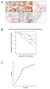
Figure 1
Figure 1
(A) Immunohistochemical staining of CIAPIN1 in CRC tissues. (a) Normal colonic epithelium, strong staining (+++) was observed in the normal colon epithelial tissues, mainly in the epithelial cells, and no evidence of expression of CIAPIN1 was noted in cells of the germinal layer (×200). (b) Gland hyperplasia of colonic mucous membrane, strong staining (+++) of CIAPIN1 was observed in dysplasia colonic epithelial tissues, there was no significant difference in the expression level of CIAPIN1 protein between normal and dysplasia colon tissues (×200). (c) Well-differentiated adenocarcinoma of colon, intermediate staining (++) of CIAPIN1 in CRC tissues with high differentiation mainly in the epithelial tissues (×200). (d) Moderately differentiated adenocarcinoma of colon, weak staining (+) of CIAPIN1 in CRC tissues with moderately differentiation, mainly in the epithelial tissues (×200). (e) Poorly differentiated adenocarcinoma of colon, negative staining (-) of CIAPIN1 in CRC tissues with poorly differentiation (×200). (f) Negative control slides using anti-His as the primary antibody. (B) Overall survival of patients determined by the immunoreactivity of CIAPIN1. Overall survival analysis using the Kaplan-Meyer method revealed that CRC patients with relatively high expression of CIAPIN1 had a more favourable prognosis compared to those with low expression (P = 0.0002). (C) Receiver operating characteristic (ROC) curve for non-local recurrence (circles) within 5 years and survival (triangles) after 5 years.
Figure 2
Figure 2
Detection of CIAPIN1 expression in freshly obtained CRC tissues Western blot analysis. (A) CIAPIN1 expression level in cancerous and adjacent normal tissues from the same patients. (B) CIAPIN1 expression level in poorly, moderately, and well-differentiated cancer tissues. (C) CIAPIN1 expression in cancer tissues with different TNM stages. β-Actin staining was used as a control for equal protein loading.
Figure 3
Figure 3
(A) RT-PCR and western blotting results showing CIAPIN1 transcripts and protein in CRC cell lines. β-Actin was used as an internal control. (B) Electron microscopy of colon cancer cells during the process of induced differentiation. (a) Undifferentiated colon cells; (b-f) Changes in ultra-microstructure during colon cell differentiation. There were special conformations such as cryptate structures, tight junctions and macula adherens between cells, as well as polarisation of microvilli in differentiated HT29 and SW480 cells. (C) AP level during induced cell differentiation in two different colon cancer cell lines. AP levels in HT29 and SW480 were measured from the 1st to the 5th day during the induced differentiation process and compared with untreated cells (day 0). (D) RT-PCR and Western blotting results showing CIAPIN1 transcripts and protein changes during HT29 and SW480 cell line differentiation from the 1st to 5th day of induced differentiation compared with untreated cells (day 0). β-Actin was used as an internal control.

Similar articles

Cited by

References

    1. Markowitz SD, Bertagnolli MM. Molecular origins of cancer: Molecular basis of colorectal cancer. N Engl J Med. 2009;361(25):2449–2460. doi: 10.1056/NEJMra0804588. - DOI - PMC - PubMed
    1. Center MM, Jemal A, Smith RA, Ward E. Worldwide variations in colorectal cancer. CA Cancer J Clin. 2009;59(6):366–378. doi: 10.3322/caac.20038. - DOI - PubMed
    1. Wang YX, Zhang XY, Zhang BF, Yang CQ, Chen XM, Gao HJ. Initial study of microRNA expression profiles of colonic cancer without lymph node metastasis. J Dig Dis. 2010;11(1):50–54. doi: 10.1111/j.1751-2980.2009.00413.x. - DOI - PubMed
    1. Punt CJ, Tol J. More is less -- combining targeted therapies in metastatic colorectal cancer. Nat Rev Clin Oncol. 2009;6(12):731–733. doi: 10.1038/nrclinonc.2009.168. - DOI - PubMed
    1. Hong Y, Downey T, Eu KW, Koh PK, Cheah PY. A 'metastasis-prone' signature for early-stage mismatch-repair proficient sporadic colorectal cancer patients and its implications for possible therapeutics. Clin Exp Metastasis. 2010;27(2):83–90. doi: 10.1007/s10585-010-9305-4. - DOI - PubMed

Publication types

MeSH terms

Substances