. 2010 Sep 5:10:96.
doi: 10.1186/1471-213X-10-96.

Mtmr8 is essential for vasculature development in zebrafish embryos

Affiliations

Mtmr8 is essential for vasculature development in zebrafish embryos

Jie Mei et al. BMC Dev Biol. .

Abstract

Background: Embryonic morphogenesis of vascular and muscular systems is tightly coordinated, and a functional cooperation of Mtmr8 with PI3K in actin filament modeling and muscle development has been revealed in zebrafish. Here, we attempt to explore the function of Mtmr8 in vasculature development parallel to its function in muscle development.

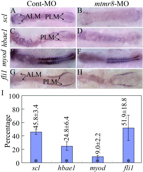
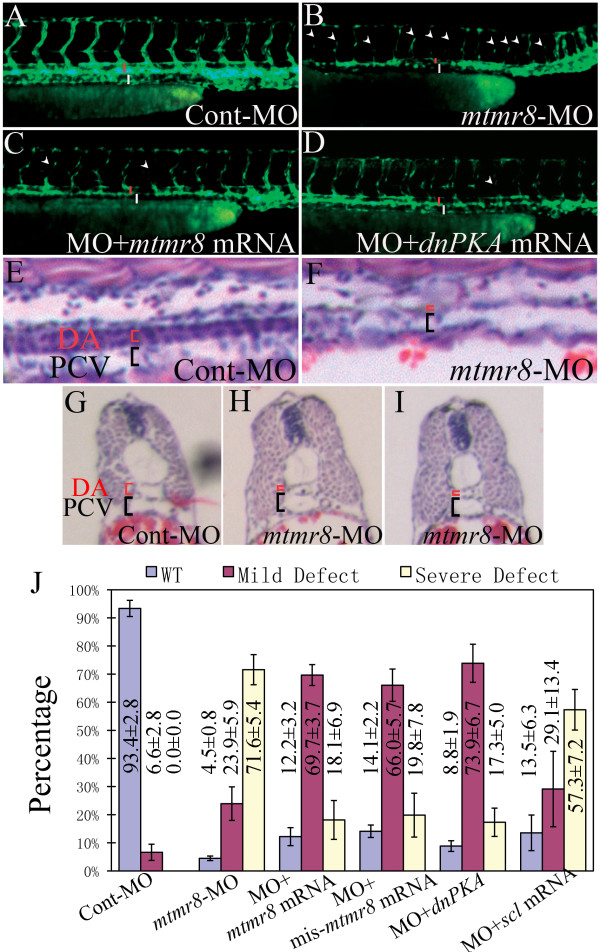
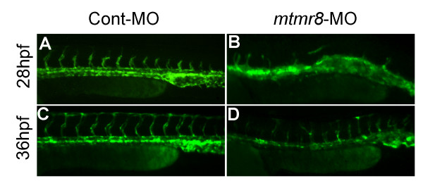
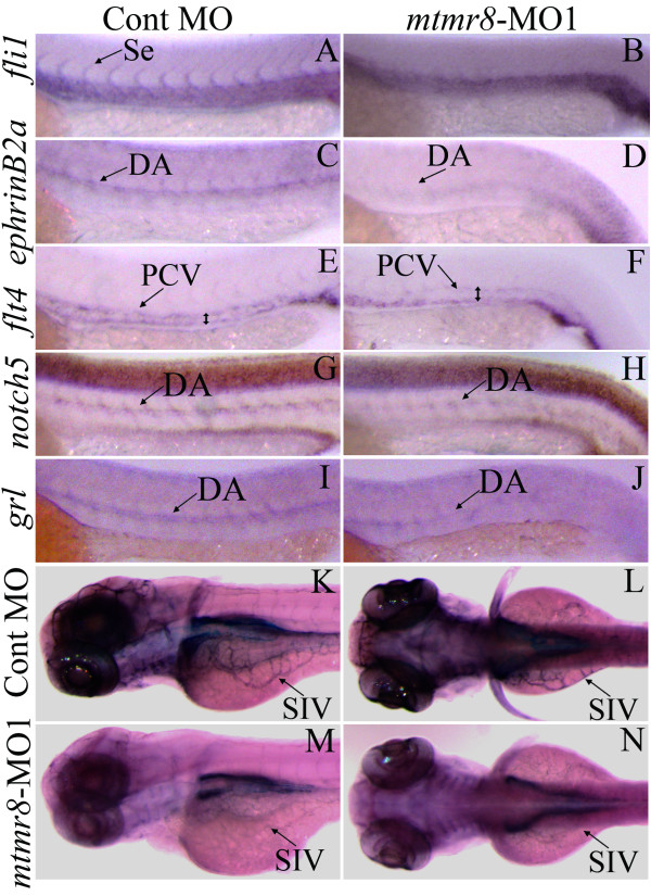
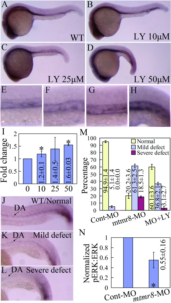
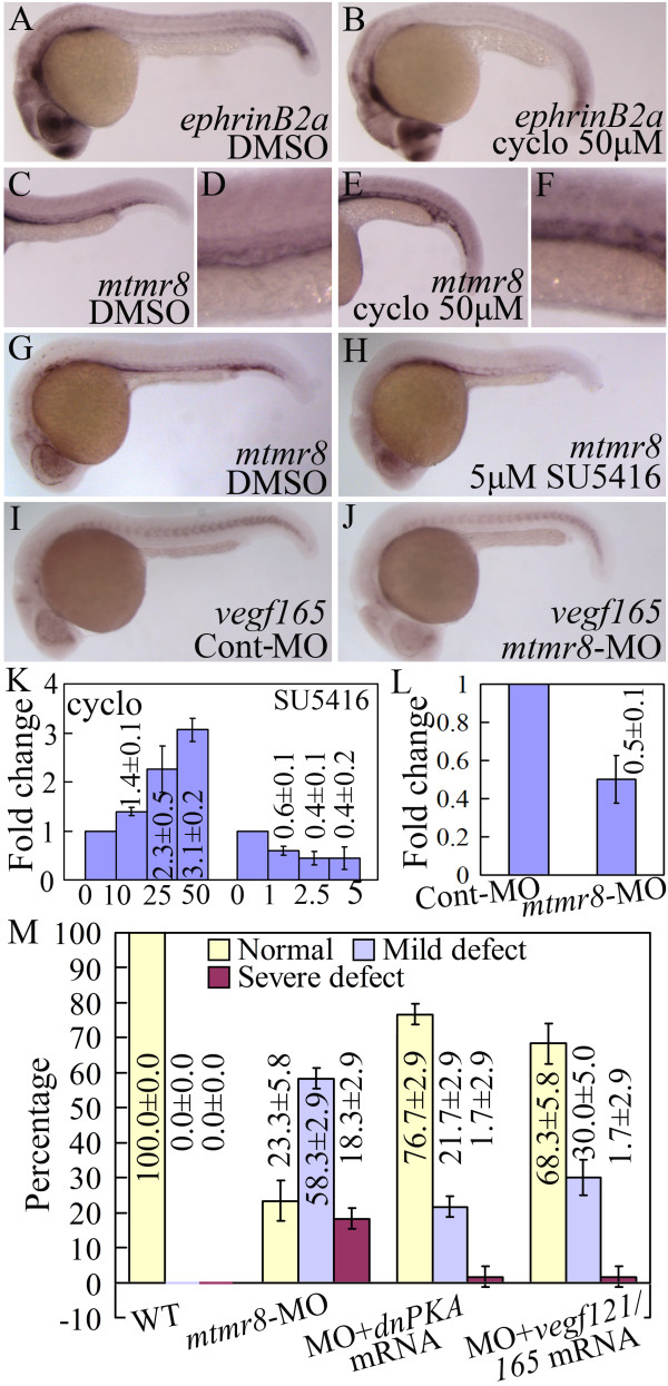
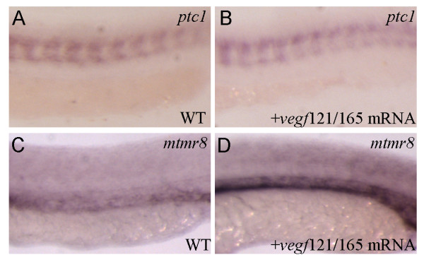
Results: During early stage of somitogenesis, mtmr8 expression was detected in both somitic mesodem and ventral mesoderm. Knockdown of mtmr8 by morpholino impairs arterial endothelial marker expression, and results in endothelial cell reduction and vasculogenesis defects, such as retardation in intersegmental vessel development and interruption of trunk dorsal aorta. Moreover, mtmr8 morphants show loss of arterial endothelial cell identity in dorsal aorta, which is effectively rescued by low concentration of PI3K inhibitor, and by over-expression of dnPKA mRNA or vegf mRNA. Interestingly, mtmr8 expression is up-regulated when zebrafish embryos are treated with specific inhibitor of Hedgehog pathway that abolishes arterial marker expression.

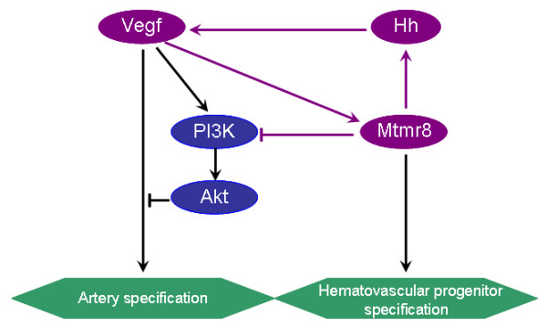
Conclusion: These data indicate that Mtmr8 is essential for vasculature development in zebrafish embryos, and may play a role in arterial specification through repressing PI3K activity. It is suggested that Mtmr8 should represent a novel element of the Hedgehog/PI3K/VEGF signaling cascade that controls arterial specification.


Figures

Figure 1
Figure 1
Early marker genes for hemato-vascular progenitors, such as early angioblast/hematopoietic mesoderm marker genes scl (A, B) and hbae1(C, D), somitic mesoderm marker gene myod(E, F), and endothelial cell marker gene fli1(G, H), are affected in lateral mesoderm of mtmr8 knockdown embryos during somitogenesis. All pictures were taken from 12 somites stage embryos. Panels are flat-mounted dorsal views (anterior toward the left). ALM (anterior lateral mesoderm) and PLM (posterior lateral mesoderm) are indicated by arrows. (I) Percentages of scl, hbae1, myod and fli1 expression of mtmr8 morphant embryos relative to Cont-MO embryos revealed by Q-RT-PCR. * indicates significance of p < 0.05.
Figure 2
Figure 2
Mtmr8 knockdown results in vascular defects. (A-D) Vascular morphology in Cont-MO (A), mtmr8-MO (B), mtmr8-MO+ mtmr8 mRNA (C) and mtmr8-MO+dnPKA mRNA Tg(kdrl:GFP)la116 embryos at 48 hpf. The presumptive lumen of DA and PCV are indicated by red and white bars, respectively. White arrowheads point to the interruption of Se. (E, F) Longitudinal sections of trunk regions in 48 hpf corresponding embryos. (G-I) Transverse sections of trunk regions in 48 hpf corresponding embryos. Sections were stained with hematoxylin and eosin. (J) Statistical data of three different defects in three independent experiments of Cont-MO, mtmr8-MO, mtmr8-MO+mtmr8 mRNA, mtmr8-MO+mis-mtmr8 mRNA, mtmr8-MO+dnPKA mRNA, and mtmr8-MO+scl mRNA.
Figure 3
Figure 3
The effects of mtmr8 knockdown on early vasculature development and blood formation in Tg(kdrl:GFP)la116 transgenic embryos. (A-D) Vascular morphology in Cont-MO (A, C) and mtmr8-MO (B, D) Tg(kdrl:GFP)la116 transgenic embryos at 28 hpf (A, B) and 36 hpf (C, D), respectively.
Figure 4
Figure 4
The effects of mtmr8 knockdown on vascular marker genes and vascular endothelium. (A-J) Whole mount in situ hybridization with the vascular marker gene-specific probes (as shown on the left) in Cont-MO (A, C, E, G and I) and mtmr8-MO (B, D, F, H and J) embryos at 26 hpf. The signals of DA, PCV and Se are indicated by arrows, and the PCV expansion size is indicated by double-arrowheads in the flt4 probe hybridization (E and F). (K-N) Lateral (K, M) and dorsal (L, N) views of alkaline phosphatase-stained Cont-MO (K, L) and mtmr8-MO (M, N) embryos at 3 dpf.
Figure 5
Figure 5
Mtmr8 negatively regulates the PI3K/Akt pathway. (A-D) Mtmr8 expression in wild type (WT) embryo (A) and the treated embryos (C-D) with different doses of LY294002 (LY) at 26 hpf. (E-H) The corresponding amplification of A-D, showing the detailed changes in the trunk vasculature. (I) Mtmr8 expression changes revealed by Q-RT-PCR in embryos treated with different doses of LY294002 (μM). (J-L) Three phenotypes of ephrinB2a expression and quantification data (M) in 26 hpf Mtmr8 morphants (30 embryos in each experiment group). (N) Quantitative Western blot data of ERK phosphorylation in 20-somite Mtmr8 morphants. * Indicates significance of p < 0.05.
Figure 6
Figure 6
(A-F) The expression of ephrinB2a (A, B) and mtmr8 (C-F) in 26 hpf embryos incubated with DMSO (A, C) or cyclopamine (B, E). (D, F) are magnification of (C, E) in the trunk vasculture. (G-H) Mtmr8 expression in 30 hpf embryos incubated with DMSO (G) or SU5416 (H). (I-J) Vegf165 expression in 26 hpf Cont-MO and Mtmr8 morphant embryos. (K) Mtmr8 expression changes revealed by Q-RT-PCR in embryos treated with different doses of SU5416 and cyclopamine (μM). (L) Vegf165 expression in 26 hpf Cont-MO and Mtmr8 morphant embryos revealed by Q-RT-PCR. (M) Statistical data of different defect phenotypes at 26 hpf in Mtmr8-MO, Mtmr8-MO+dnPKA mRNA, and Mtmr8-MO+vegf121/165 mRNA embryos.
Figure 7
Figure 7
The effect of Vegf overexpression on mtmr8 expression. (A, B) Expression of ptc1 in wild type (WT) embryo (A) and in vegf121/165 mRNA injected embryo (B). (C, D) Expression of mtmr8 in wild type (WT) embryo (C) and in vegf121/165 mRNA injected embryo (D). All embryos in 25 hpf; anterior toward the left.
Figure 8
Figure 8
A hypothesized signal pathway that Mtmr8 regulates artery specification and hematovascular progenitor specification in zebrafish embryo. Mtmr8 inhibits PI3K/Akt pathway, and activates Hh and Vegf signaling pathway, which may interact with each other in regulating vasculature development. The pink arrows indicate the regulatory loop.

Similar articles

Cited by

References

    1. Hunter T. Anti-phosphatases take the stage. Nat Genet. 1998;18:303–5. doi: 10.1038/ng0498-303. - DOI - PubMed
    1. Jiang BH, Zheng JZ, Aoki M, Vogt PK. Phosphatidylinositol 3-kinase signaling mediates angiogenesis and expression of vascular endothelial growth factor in endothelial cells. Proc Natl Acad Sci USA. 2000;97:1749–53. doi: 10.1073/pnas.040560897. - DOI - PMC - PubMed
    1. Wen S, Stolarov J, Myers MP, Su JD, Wigler MH, Tonks NK, Durden DL. PTEN controls tumor-induced angiogenesis. Proc Natl Acad Sci USA. 2001;98:4622–7. doi: 10.1073/pnas.081063798. - DOI - PMC - PubMed
    1. Hu Z, Lee IH, Wang X, Sheng H, Zhang L, Du J, Mitch WE. PTEN expression contributes to the regulation of muscle protein degradation in diabetes. Diabetes. 2007;56:2449–56. doi: 10.2337/db06-1731. - DOI - PubMed
    1. Pramanik K, Chun CZ, Garnaas MK, Samant GV, Li K, Horswill MA, North PE, Ramchandran R. Dusp-5 and Snrk-1 coordinately function during vascular development and disease. Blood. 2009;113:1184–91. doi: 10.1182/blood-2008-06-162180. - DOI - PMC - PubMed

Publication types

MeSH terms

Substances

LinkOut - more resources