. 2010 Sep 8;99(5):1408-15.
doi: 10.1016/j.bpj.2010.06.042.

So little source, so much sink: requirements for afterdepolarizations to propagate in tissue

Affiliations

So little source, so much sink: requirements for afterdepolarizations to propagate in tissue

Yuanfang Xie et al. Biophys J. .

Abstract

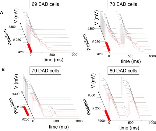
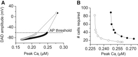
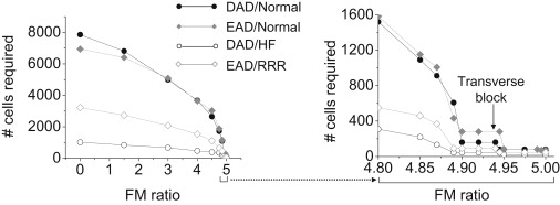
How early (EADs) and delayed afterdepolarizations (DADs) overcome electrotonic source-sink mismatches in tissue to trigger premature ventricular complexes remains incompletely understood. To study this question, we used a rabbit ventricular action potential model to simulate tissues in which a central area of contiguous myocytes susceptible to EADs or DADs was surrounded by unsusceptible tissue. In 1D tissue with normal longitudinal conduction velocity (0.55 m/s), the numbers of contiguous susceptible myocytes required for an EAD and a barely suprathreshold DAD to trigger a propagating action potential were 70 and 80, respectively. In 2D tissue, these numbers increased to 6940 and 7854, and in 3D tissue to 696,910 and 817,280. These numbers were significantly decreased by reduced gap junction conductance, simulated fibrosis, reduced repolarization reserve and heart failure electrical remodeling. In conclusion, the source-sink mismatch in well-coupled cardiac tissue powerfully protects the heart from arrhythmias due to sporadic afterdepolarizations. Structural and electrophysiological remodeling decrease these numbers significantly but still require synchronization mechanisms for EADs and DADs to overcome the robust protective effects of source-sink mismatch.


Figures

Figure 1
Figure 1
(A) Superimposed AP traces corresponding to the normal rabbit ventricular cell AP model (black line), the cell AP model with reduced repolarization reserve (RRR, blue line), and the cell model with an EAD triggering a spontaneous AP (red line). All traces were obtained from a single uncoupled cell paced at a PCL of 600 ms. (B) Superimposed AP traces corresponding to normal (black line) and heart failure (red line) parameters in rabbit ventricular cell AP models, in which the paced AP (PCL 500 ms) is followed by a DAD, which triggers a second AP. (Inset) Corresponding DAD amplitudes when the Na and Ca currents are blocked after the first paced AP. (C) Schematic of 1D tissue, with central region of EAD/DAD susceptible cells demarcated in red, and unsusceptible cells in blue. (D) Schematic of 2D tissue, with central elliptical region of EAD-/DAD-susceptible cells demarcated in red. (E) Schematic of bricklike 2D tissue with fibroblasts (F) randomly interspersed at the ends (I) or sides (II) of the myocytes (M).
Figure 2
Figure 2
(A) Selected AP traces along a 1D cable, with the red traces indicating the EAD-susceptible cells in the central region, and the normal unsusceptible cells in black. The EAD failed to propagate with 69 susceptible cells in the central region (left), but propagated successfully with 70 susceptible cells (right). (B) Same as for A, but with the central region exhibiting DAD-susceptible cells. The DAD failed to trigger an AP with 79 susceptible (B) cells in the central region (left), but did so successfully with 80 susceptible cells (right). Gap junction conductance was 780 nS.
Figure 3
Figure 3
(A) DAD amplitude as a function of the total amount of SR Ca release in a single myocyte. A larger total amount of SR Ca release is required for the DAD to reach threshold when the SR Ca release time constant was increased from τ1 = 10 ms (open circles) to 50 ms (solid circles). (B) Number of contiguous DAD-susceptible myocytes in the central region of a 6-cm 1D cable required for the DAD to trigger a PVC versus the total amount of SR Ca released for a normal (open circles, τ1 = 10 ms) versus a slowed (solid circles, τ1 = 50 ms) SR Ca release time constant.
Figure 4
Figure 4
Number of contiguous susceptible myocytes required to trigger an EAD- or DAD-mediated PVC in simulated 2D tissue with lateral fibrosis. Fibroblasts were randomly interspersed exclusively along the sides of myocytes throughout the entire tissue (Fig. 1E, II), with EAD- or DAD-susceptible myocytes only in the central region. As the fibroblast/myocyte (FM) ratio increased, the required size of the central region (and hence the number of susceptible cells in the central region) progressively decreased, approaching the 1D case at the maximum FM ratio of ∼5, above which transverse propagation failed. The expanded region of FM ratio 4.8–5.0 is shown at right. EAD/Normal refers to EAD-susceptible cells in the central region surrounded by nonsusceptible normal cells. The other labels can be interpreted similarly, where the surrounding tissue was either normal or exhibited RRR or HF changes. Gap junction conductance (Ggap) is 780 nS in all cases.

References

    1. Joyner R.W., Sugiura H., Tan R.C. Unidirectional block between isolated rabbit ventricular cells coupled by a variable resistance. Biophys. J. 1991;60:1038–1045. - PMC - PubMed
    1. Shaw R.M., Rudy Y. Ionic mechanisms of propagation in cardiac tissue. Roles of the sodium and L-type calcium currents during reduced excitability and decreased gap junction coupling. Circ. Res. 1997;81:727–741. - PubMed
    1. Spach M.S., Boineau J.P. Microfibrosis produces electrical load variations due to loss of side-to-side cell connections: a major mechanism of structural heart disease arrhythmias. Pacing Clin. Electrophysiol. 1997;20:397–413. - PubMed
    1. Rohr S., Kucera J.P., Kléber A.G. Paradoxical improvement of impulse conduction in cardiac tissue by partial cellular uncoupling. Science. 1997;275:841–844. - PubMed
    1. Wang Y.G., Kumar R., Joyner R.W. Electrical interactions between a real ventricular cell and an anisotropic two-dimensional sheet of model cells. Am. J. Physiol. Heart Circ. Physiol. 2000;278:H452–H460. - PubMed

Publication types