Review
. 2010:92:73-129.
doi: 10.1016/S0070-2153(10)92003-6.

Canonical and non-canonical Notch ligands

Affiliations
Review

Canonical and non-canonical Notch ligands

Brendan D'Souza et al. Curr Top Dev Biol. 2010.

Abstract

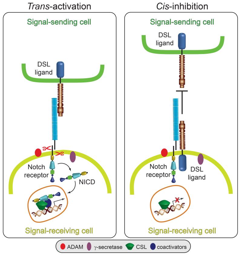
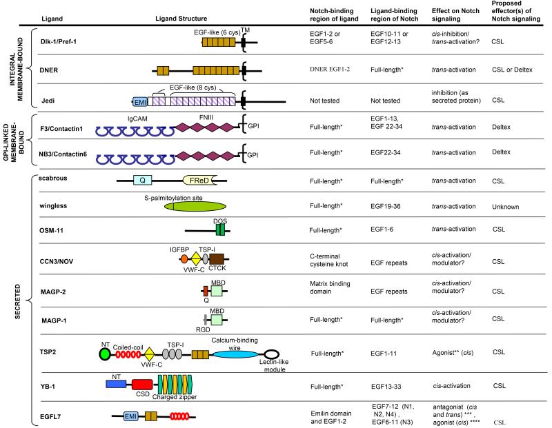
Notch signaling induced by canonical Notch ligands is critical for normal embryonic development and tissue homeostasis through the regulation of a variety of cell fate decisions and cellular processes. Activation of Notch signaling is normally tightly controlled by direct interactions with ligand-expressing cells, and dysregulated Notch signaling is associated with developmental abnormalities and cancer. While canonical Notch ligands are responsible for the majority of Notch signaling, a diverse group of structurally unrelated noncanonical ligands has also been identified that activate Notch and likely contribute to the pleiotropic effects of Notch signaling. Soluble forms of both canonical and noncanonical ligands have been isolated, some of which block Notch signaling and could serve as natural inhibitors of this pathway. Ligand activity can also be indirectly regulated by other signaling pathways at the level of ligand expression, serving to spatiotemporally compartmentalize Notch signaling activity and integrate Notch signaling into a molecular network that orchestrates developmental events. Here, we review the molecular mechanisms underlying the dual role of Notch ligands as activators and inhibitors of Notch signaling. Additionally, evidence that Notch ligands function independent of Notch is presented. We also discuss how ligand posttranslational modification, endocytosis, proteolysis, and spatiotemporal expression regulate their signaling activity.


Figures

Fig. 1
Fig. 1. Models for DSL ligand trans-activation and cis-inhibition in Notch signaling
Ligand expressed on the surface of the signal-sending cell binds to Notch expressed on the surface of the signal-receiving cell (trans-interactions) and induces sequential cleavages by A-Disintegrin-And-Metalloprotease (ADAM) and -secretase in Notch releasing the Notch intracellular domain (NICD) from the membrane. NICD translocates to the nucleus where it directly interacts with the CSL (CBF1, Su(H), LAG1) transcription factor and recruits coactivators to induce Notch target gene expression. Ligand binding to Notch expressed in the same cell (cis-interactions) prevents Notch activation by trans- ligand by competing with trans-ligand for Notch binding.
Fig. 2
Fig. 2. Structural domains of canonical ligands
The extracellular domains of canonical ligands are characterized by the presence of an N-terminal (NT) domain followed by a Delta/Serrate/LAG-2 (DSL) domain and multiple tandemly arranged Epidermal Growth Factor (EGF)-like repeats (see text for details). The DSL domain together with the flanking NT domain and the first two EGF repeats containing the Delta and OSM-11-like proteins (DOS) motif are required for canonical ligands to bind Notch. The NT domain of vertebrate and Drosophila ligands is subdivided into a region containing six conserved cysteine residues, N1 and a cysteine- free region, N2. Serrate/Jagged ligands contain an additional cysteine-rich region not present in Delta-like ligands. The intracellular domains of some canonical ligands contain a carboxy-terminal PSD-95/Dlg/ZO-1-ligand (PDZL) motif that plays a role independent of Notch signaling. C. elegans DSL ligands lack a DOS motif but have been proposed to cooperate with DOS-only containing ligands (not depicted) to activate Notch signaling. Dll3 is the most structurally divergent vertebrate DSL ligand and lacks structural features required by other DSL ligands to bind and activate Notch.
Fig. 3
Fig. 3. Models for distinct endocytic events by the ligand cell to activate signaling in the Notch cell
Prior to Notch engagement, endocytosis allows ligand to enter the sorting endosome (SE) or recycling endosome (RE) where it is processed into an active ligand and returned to the cell surface to activate Notch. Ligand ubiquitination by Mib may facilitate interactions with epsin that direct the required endocytosis and/or trafficking. Alternatively, ligand binding to Notch may induce ligand ubiquitination for recruitment of epsin to orchestrate the formation of a clathrin- coated endocytic structure specialized in force generation to pull the non- covalent heterodimeric Notch apart. Heterodimer dissociation would account for the observed uptake of the Notch extracellular domain (NECD) by ligand cells. In the early endosome (EE), internalized NECD dissociates from the ligand and trafficks to the late endsome (LE) where it is targeted for lysosomal degradation. Ligand dissociated in the EE traffics to the SE or RE for return to the cell surface where it is available to activate Notch on adjacent cells. Removal of the NECD exposes the ADAM site in the membrane-bound heterodimer subunit to facilitate γ-secretase cleavage and release of the Notch intracellular domain (NICD) from the membrane. Released NICD translocates to nucleus where it interacts with CSL to activate Notch target gene transcription. As discussed in the text, these models that account for the critical requirement for endocytosis by the ligand cell to activate signaling in the Notch cell may not be mutually exclusive.
Fig. 4
Fig. 4. Regulation of DSL ligand signaling activity by proteolysis
Mammalian and Drosophila DSL ligands undergo proteolytic cleavages within the juxtamembrane and intramembrane regions. A-Disintegrin-And-Metalloprotease (ADAM) mediated cleavage (1) of mammalian and Drosophila DSL (Delta/Serrate/LAG-2) ligands within the juxtamembrane region results in shedding of the extracellular domain (2, ECD). The shed ECD requires clustering to activate Notch signaling (3). Although unclustered soluble ECD can bind Notch, it may antagonize Notch signaling (4). In mammalian cells, the remaining membrane-tethered ADAM cleavage product, the membrane-tethered fragment containing the intracellular domain (TMICD, 5) may undergo further cleavage by -secretase (6) to release the intracellular domain (ICD) from the membrane (7) allowing it translocate to the nucleus and activate gene transcription (8) (see text for details). However, the Drosophila Delta (dDelta) TMICD (5) is not further processed and could antagonize Notch signaling (see text for details). Like mammalian DSL ligands, dDelta also undergoes intramembrane cleavage, however, this event does not require prior ADAM cleavage and is catalyzed by a thiol-sensitive activity (TSA, 9). It is unclear if the resulting cleavage products remain membrane-tethered. If the ECD containing fragment (ECDTM) remains membrane-tethered (10), it could antagonize Notch signaling, but if released from the membrane, ECDTM could function as proposed for soluble ECD (2, 3, 4) (see text for details). If the ICD-containing intramembrane cleavage product TMICDTSA remains membrane-bound (11), it could antagonize Notch signaling, but if released from the membrane (7), TMICDTSA could translocate to the nucleus and activate gene transcription (8) (see text for details).
Fig. 5
Fig. 5. Non-canonical ligand structure and proposed effects on Notch signaling
Non-canonical ligands lack a DSL domain (Delta/Serrate/LAG-2), are structurally diverse and include integral- and GPI-linked membrane proteins as well as secreted proteins (see text for details). EGF-like (6 cys), 6-cysteine epidermal growth factor-like repeat as found in canonical ligands; cys, cysteine; TM, transmembrane domain, CSL (CBF1, Su(H), LAG1); EMI, emilin-like domain; EGF-like (8 cys), EGF-like motif with 8 cysteines that is not laminin-like; Ig-CAM, immunoglobulin-containing cell adhesion molecule domain; FNIII, fibronectin type III domain; GPI, glycosylphosphatidylinositol; Q, glutamine-rich region; FReD, fibrinogen-related domain; DOS, Delta and OSM-11-like proteins; IGFBP, insulin-like growth factor-binding protein-like domain; VWF-C, von Willebrand factor type C-like domain; TSP-1, thrombospondin type 1-like domain; CTCK, C-terminal cysteine knot domain; MBD, matrix binding domain; RGD, integrin binding motif; NT, N-terminal domain; CSD, cold shock domain, N1, Notch1; N2, Notch2; N3, Notch3; N4, Notch4. *Only full-length constructs were tested for binding. **Agonist of Jagged1 signaling ***Antagonist of Jagged1 signaling. **** Agonist of Dll4 (Delta-like 4) signaling.

References

    1. Ahimou F, Mok LP, Bardot B, Wesley C. The adhesion force of Notch with Delta and the rate of Notch signaling. J Cell Biol. 2004;167:1217–1229. - PMC - PubMed
    1. Aho S. Soluble form of Jagged1: unique product of epithelial keratinocytes and a regulator of keratinocyte differentiation. J Cell Biochem. 2004;92:1271–1281. - PubMed
    1. Akai J, Halley PA, Storey KG. FGF-dependent Notch signaling maintains the spinal cord stem zone. Genes Dev. 2005;19:2877–2887. - PMC - PubMed
    1. Albig AR, Becenti DJ, Roy TG, Schiemann WP. Microfibril-associate glycoprotein-2 (MAGP-2) promotes angiogenic cell sprouting by blocking notch signaling in endothelial cells. Microvasc Res. 2008 - PMC - PubMed
    1. Amsen D, Blander JM, Lee GR, Tanigaki K, Honjo T, Flavell RA. Instruction of distinct CD4 T helper cell fates by different notch ligands on antigen-presenting cells. Cell. 2004;117:515–526. - PubMed

Publication types

LinkOut - more resources