Area postrema projects to FoxP2 neurons of the pre-locus coeruleus and parabrachial nuclei: brainstem sites implicated in sodium appetite regulation
- PMID: 20816675
- PMCID: PMC2955772
- DOI: 10.1016/j.brainres.2010.08.085
Area postrema projects to FoxP2 neurons of the pre-locus coeruleus and parabrachial nuclei: brainstem sites implicated in sodium appetite regulation
Abstract
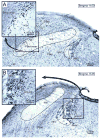
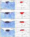
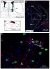
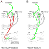
The area postrema (AP) is a circumventricular organ located in the dorsal midline of the medulla. It functions as a chemosensor for blood-borne peptides and solutes, and converts this information into neural signals that are transmitted to the nucleus tractus solitarius (NTS) and parabrachial nucleus (PB). One of its NTS targets in the rat is the aldosterone-sensitive neurons which contain the enzyme 11 β-hydroxysteroid dehydrogenase type 2 (HSD2). The HSD2 neurons are part of a central network involved in sodium appetite regulation, and they innervate numerous brain sites including the pre-locus coeruleus (pre-LC) and PB external lateral-inner (PBel-inner) cell groups of the dorsolateral pons. Both pontine cell groups express the transcription factor FoxP2 and become c-Fos activated following sodium depletion. Because the AP is a component in this network, we wanted to determine whether it also projects to the same sites as the HSD2 neurons. By using a combination of anterograde axonal and retrograde cell body tract-tracing techniques in individual rats, we show that the AP projects to FoxP2 immunoreactive neurons in the pre-LC and PBel-inner. Thus, the AP sends a direct projection to both the first-order medullary (HSD2 neurons of the NTS) and the second-order dorsolateral pontine neurons (pre-LC and PB-el inner neurons). All three sites transmit information related to systemic sodium depletion to forebrain sites and are part of the central neural circuitry that regulates the complex behavior of sodium appetite.
Copyright © 2010 Elsevier B.V. All rights reserved.
Figures


Similar articles
-
FoxP2 brainstem neurons project to sodium appetite regulatory sites.J Chem Neuroanat. 2011 Sep;42(1):1-23. doi: 10.1016/j.jchemneu.2011.05.003. Epub 2011 May 13. J Chem Neuroanat. 2011. PMID: 21605659 Free PMC article. Review.
-
Serotonergic inputs to FoxP2 neurons of the pre-locus coeruleus and parabrachial nuclei that project to the ventral tegmental area.Neuroscience. 2011 Oct 13;193:229-40. doi: 10.1016/j.neuroscience.2011.07.008. Epub 2011 Jul 18. Neuroscience. 2011. PMID: 21784133 Free PMC article.
-
Aldosterone-sensitive neurons in the nucleus of the solitary: efferent projections.J Comp Neurol. 2006 Sep 20;498(3):223-50. J Comp Neurol. 2006. PMID: 16933386
-
Sodium deprivation and salt intake activate separate neuronal subpopulations in the nucleus of the solitary tract and the parabrachial complex.J Comp Neurol. 2007 Oct 1;504(4):379-403. doi: 10.1002/cne.21452. J Comp Neurol. 2007. PMID: 17663450
-
Control of sodium appetite by hindbrain aldosterone-sensitive neurons.Mol Cell Endocrinol. 2024 Oct 1;592:112323. doi: 10.1016/j.mce.2024.112323. Epub 2024 Jun 26. Mol Cell Endocrinol. 2024. PMID: 38936597 Review.
Cited by
-
FoxP2 brainstem neurons project to sodium appetite regulatory sites.J Chem Neuroanat. 2011 Sep;42(1):1-23. doi: 10.1016/j.jchemneu.2011.05.003. Epub 2011 May 13. J Chem Neuroanat. 2011. PMID: 21605659 Free PMC article. Review.
-
Transcription factors define the neuroanatomical organization of the medullary reticular formation.Front Neuroanat. 2013 May 14;7:7. doi: 10.3389/fnana.2013.00007. eCollection 2013. Front Neuroanat. 2013. PMID: 23717265 Free PMC article.
-
The Expression Pattern of the Na(+) Sensor, Na(X) in the Hydromineral Homeostatic Network: A Comparative Study between the Rat and Mouse.Front Neuroanat. 2012 Jul 19;6:26. doi: 10.3389/fnana.2012.00026. eCollection 2012. Front Neuroanat. 2012. PMID: 22833716 Free PMC article.
-
Serotonergic inputs to FoxP2 neurons of the pre-locus coeruleus and parabrachial nuclei that project to the ventral tegmental area.Neuroscience. 2011 Oct 13;193:229-40. doi: 10.1016/j.neuroscience.2011.07.008. Epub 2011 Jul 18. Neuroscience. 2011. PMID: 21784133 Free PMC article.
-
ENaC-expressing neurons in the sensory circumventricular organs become c-Fos activated following systemic sodium changes.Am J Physiol Regul Integr Comp Physiol. 2013 Nov 15;305(10):R1141-52. doi: 10.1152/ajpregu.00242.2013. Epub 2013 Sep 18. Am J Physiol Regul Integr Comp Physiol. 2013. PMID: 24049115 Free PMC article.
References
-
- Alden M, Besson JM, Bernard JF. Organization of the efferent projections from the pontine parabrachial area to the bed nucleus of the stria terminalis and neighboring regions: a PHA-L study in the rat. J Comp Neurol. 1994;341:289–314. - PubMed
-
- Bernard JF, Alden M, Besson JM. The organization of the efferent projections from the pontine parabrachial area to the amygdaloid complex: a Phaseolus vulgaris leucoagglutinin (PHA-L) study in the rat. J Comp Neurol. 1993;329:201–229. - PubMed
-
- Berthoud HR, Neuhuber WL. Functional and chemical anatomy of the afferent vagal system. Auton Neurosci. 2000;85:1–17. - PubMed
-
- Borison HL, Brizzee KR. Morphology of emetic chemoreceptor trigger zone in cat medulla oblongata. Proc Soc Exp Biol Med. 1951;77:38–42. - PubMed
-
- Contreras RJ, Stetson PW. Changes in salt intake lesions of the area postrema and the nucleus of the solitary tract in rats. Brain Res. 1981;211:355–366. - PubMed
Publication types
MeSH terms
Substances
Grants and funding
LinkOut - more resources
Full Text Sources
Other Literature Sources

